Copyright
©2012 Baishideng Publishing Group Co.
World J Gastroenterol. Jul 21, 2012; 18(27): 3623-3626
Published online Jul 21, 2012. doi: 10.3748/wjg.v18.i27.3623
Published online Jul 21, 2012. doi: 10.3748/wjg.v18.i27.3623
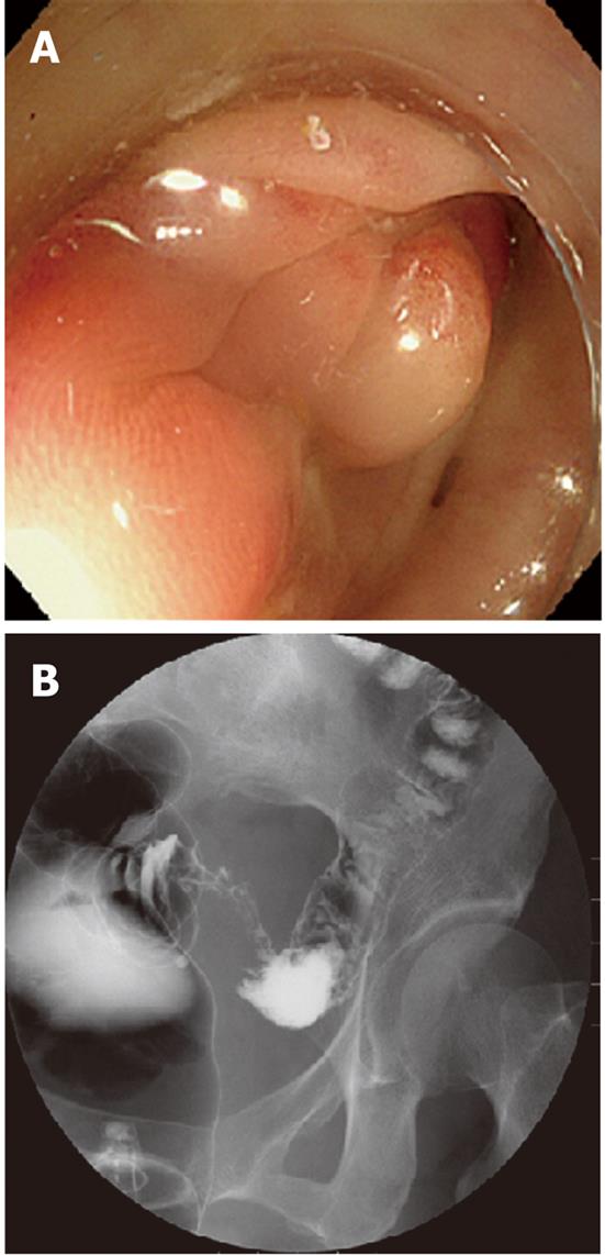
Figure 1 Colonofiberscopy and Barium enema revealed a marked, hourglass-shaped, 2-cm circumferential stenosis in the sigmoid colon.
A: Colonofiberscopy; B: Barium enema.
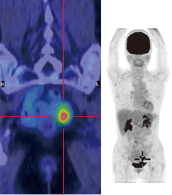
Figure 2 Fluorodeoxyglucose-positron emission tomography computed tomography imaging.
A fluorodeoxyglucose-positron emission tomography computed tomography scan showing increased fluorodeoxyglucose (FDG) uptake in the affected portion of the sigmoid colon (maximum standardized uptake value: 5.6 in the early phase and 7.4 in the late phase), but no increase in FDG uptake was observed in the surrounding lymph nodes or distant organs.
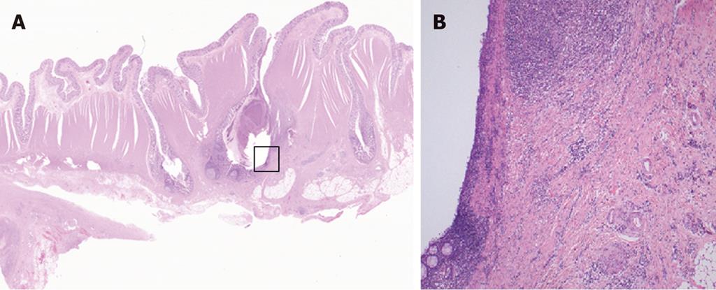
Figure 3 Magnified pathological image of resected specimen.
A: A magnified pathological image of the resected specimen reveals some diverticula in the sigmoid colon (HE stain, 10 ×); B: A magnified pathological image of the marked square in Figure 4A (diverticulosis) shows active inflammation characterized by a marked thickening of the muscularis propria, fibrous thickening of interstitial tissue, and a marked infiltration of inflammatory cells with no evidence of malignancy (100 ×).
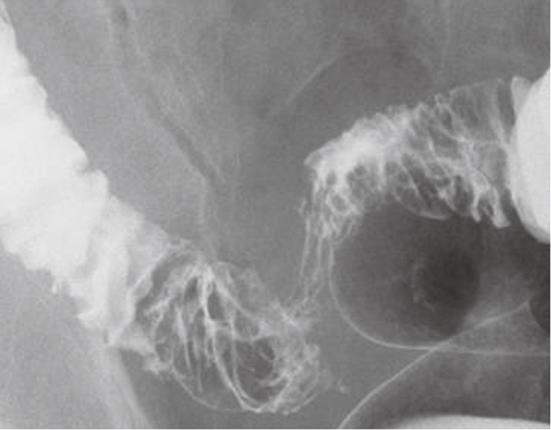
Figure 4 Barium enema revealed a segmental stenosis approximately 10 cm long, mucosal wall thickening, and cobblestone-like mucosa in the sigmoid colon.
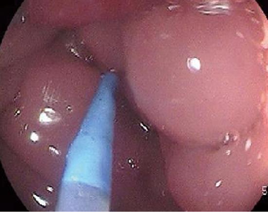
Figure 5 Colonofiberscopy detected severe stenosis in the sigmoid colon approximately 25 cm from the dentate line, and the scope could not be advanced further.
Mucosal surfaces in the visible range were edematous and red with a partial polyposis-like appearance.
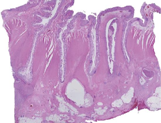
Figure 6 Pathological examination of the resected specimen.
Pathological examination of the resected specimen revealed no change in the mucosal epithelial surface, but multiple diverticula penetrated the muscularis propria with a marked thickening of the muscularis propria, fibrous thickening of interstitial tissue, severe infiltration of inflammatory cells, and a cystic change that contained inflammatory exudate and abscess formation.
- Citation: Nishiyama N, Mori H, Kobara H, Rafiq K, Fujihara S, Kobayashi M, Masaki T. Difficulty in differentiating two cases of sigmoid stenosis by diverticulitis from cancer. World J Gastroenterol 2012; 18(27): 3623-3626
- URL: https://www.wjgnet.com/1007-9327/full/v18/i27/3623.htm
- DOI: https://dx.doi.org/10.3748/wjg.v18.i27.3623


