Copyright
©2012 Baishideng Publishing Group Co.
World J Gastroenterol. Jul 14, 2012; 18(26): 3375-3378
Published online Jul 14, 2012. doi: 10.3748/wjg.v18.i26.3375
Published online Jul 14, 2012. doi: 10.3748/wjg.v18.i26.3375
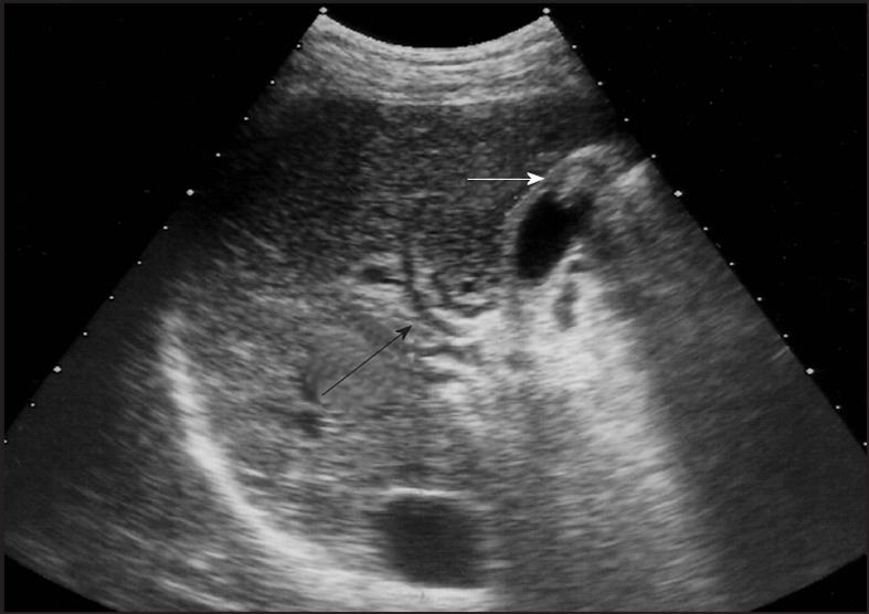
Figure 1 Liver ultrasound image showing stone (white arrow) and portal vein cavernomatous transformation (black arrow) in gallbladder.
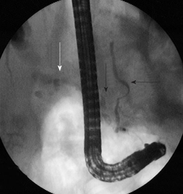
Figure 2 “Portal double ductopathy” sign with stones.
Endoscopic retrograde cholangiopancreatography image of a patient with chronic portal vein cavernomatous transformation showing irregular pancreatic duct and biliary ducts (black arrows). Main bile duct shows local stricture and dilations with cholelithiasis (white arrow).
- Citation: Harmanci O, Bayraktar Y. How can portal vein cavernous transformation cause chronic incomplete biliary obstruction? World J Gastroenterol 2012; 18(26): 3375-3378
- URL: https://www.wjgnet.com/1007-9327/full/v18/i26/3375.htm
- DOI: https://dx.doi.org/10.3748/wjg.v18.i26.3375


