Copyright
©2012 Baishideng Publishing Group Co.
World J Gastroenterol. Jul 7, 2012; 18(25): 3254-3259
Published online Jul 7, 2012. doi: 10.3748/wjg.v18.i25.3254
Published online Jul 7, 2012. doi: 10.3748/wjg.v18.i25.3254
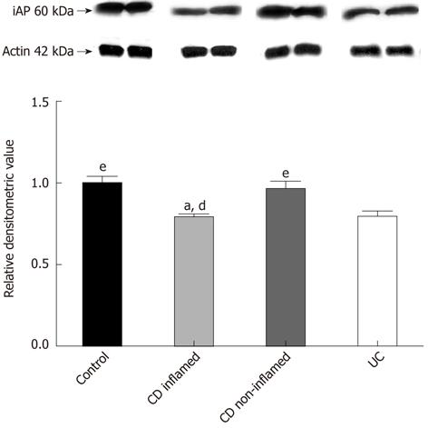
Figure 1 Protein levels of intestinal alkaline phosphatase in the colonic mucosa of children with newly diagnosed Crohn’s disease, ulcerative colitis and controls.
A: Western blotting analysis of the colonic biopsy specimens using intestinal alkaline phosphatase (iAP)-specific antibody reveals one distinct band at 60 kDa; B: Data for protein levels of iAP were obtained by computerized analysis of the Western blottings and expressed as median interquartile range. Analysis of significance was performed by the Mann-Whitney U test. aP < 0.05 vs control; dP < 0.01 vs non-inflamed CD; eP < 0.05 vs UC. CD: Crohn’s disease; UC: Ulcerative colitis.
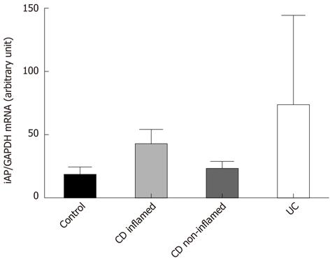
Figure 2 Intestinal alkaline phosphatase mRNA expression in the colonic mucosa of children with newly diagnosed Crohn’s disease, ulcerative colitis and controls.
iAP mRNA expression data were obtained by computerized analysis of PCR products. Optical density was corrected according to that of GAPDH. Data are expressed as median interquartile range. Analysis of significance was performed by Mann-Whitney U test. GAPDH: Glyceraldehyde-3-phosphate dehydrogenase; PCR: Polymerase chain reaction; iAP: Intestinal alkaline phosphatase; CD: Crohn’s disease; UC: Ulcerative colitis.
Figure 3 Localization of intestinal alkaline phosphatase and Toll-like receptor-4 in colon of Crohn’s disease.
Immunofluorescent staining for intestinal alkaline phosphatase (iAP) (red) and Toll-like receptor-4 (TLR4) (green) staining was performed in inflamed colon of patient with Crohn’s disease. Yellow color (merge) indicates co-localization of iAP and TLR4. For the observation of non-labeled tissues, differential interference contrast was used. Nuclei are stained with blue.
- Citation: Molnár K, Vannay &, Szebeni B, Bánki NF, Sziksz E, Cseh &, Győrffy H, Lakatos PL, Papp M, Arató A, Veres G. Intestinal alkaline phosphatase in the colonic mucosa of children with inflammatory bowel disease. World J Gastroenterol 2012; 18(25): 3254-3259
- URL: https://www.wjgnet.com/1007-9327/full/v18/i25/3254.htm
- DOI: https://dx.doi.org/10.3748/wjg.v18.i25.3254


