Copyright
©2012 Baishideng Publishing Group Co.
World J Gastroenterol. May 28, 2012; 18(20): 2576-2581
Published online May 28, 2012. doi: 10.3748/wjg.v18.i20.2576
Published online May 28, 2012. doi: 10.3748/wjg.v18.i20.2576
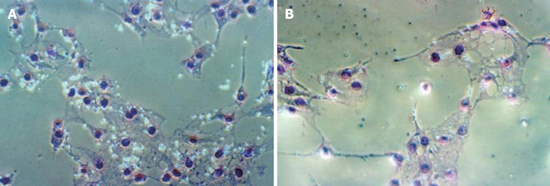
Figure 1 Effects of glycine on phagocytosis by Kupffer cells in vitro.
A: Phagotosis by Kupffer cells in the control group 30 min after the addition of latex beads, 200×; B: Phagotosis by Kupffer cells in group G3 30 min after the addition of latex beads, 200×.
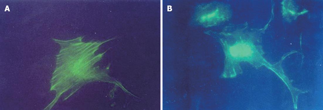
Figure 2 Effects of glycine on expression of microfilaments by Kupffer cells in vitro.
A: The expression of microfilaments by Kupffer cells in the control group, stained with Phalloidin-fluorescein isothiocyanate (FITC), 400×; B: The expression of microfilaments by Kupffer cells in group G3, stained with Phalloidin-FITC, 400×.
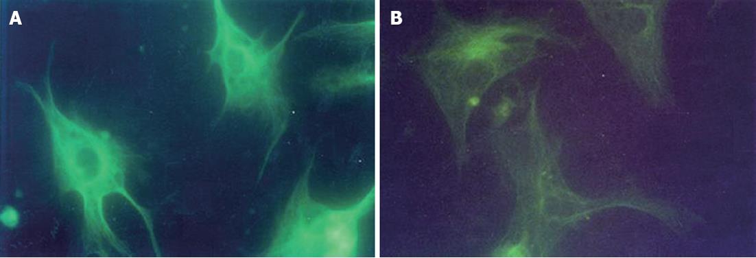
Figure 3 Effects of glycine on expression of microtubules by Kupffer cells in vitro.
A: The expression of microtubules by Kupffer cells in the control group, stained with monoclonal anti-α tubulin-fluorescein isothiocyanate (FITC) conjugate, 400×; B: The expression of microtubules by Kupffer cells in group G3, stained with monoclonal anti-α tubulin-FITC conjugate, 400×.
-
Citation: Wu HW, Yun KM, Han DW, Xu RL, Zhao YC. Effects of glycine on phagocytosis and secretion by Kupffer cells
in vitro . World J Gastroenterol 2012; 18(20): 2576-2581 - URL: https://www.wjgnet.com/1007-9327/full/v18/i20/2576.htm
- DOI: https://dx.doi.org/10.3748/wjg.v18.i20.2576


