Copyright
©2012 Baishideng Publishing Group Co.
World J Gastroenterol. Jan 14, 2012; 18(2): 150-155
Published online Jan 14, 2012. doi: 10.3748/wjg.v18.i2.150
Published online Jan 14, 2012. doi: 10.3748/wjg.v18.i2.150
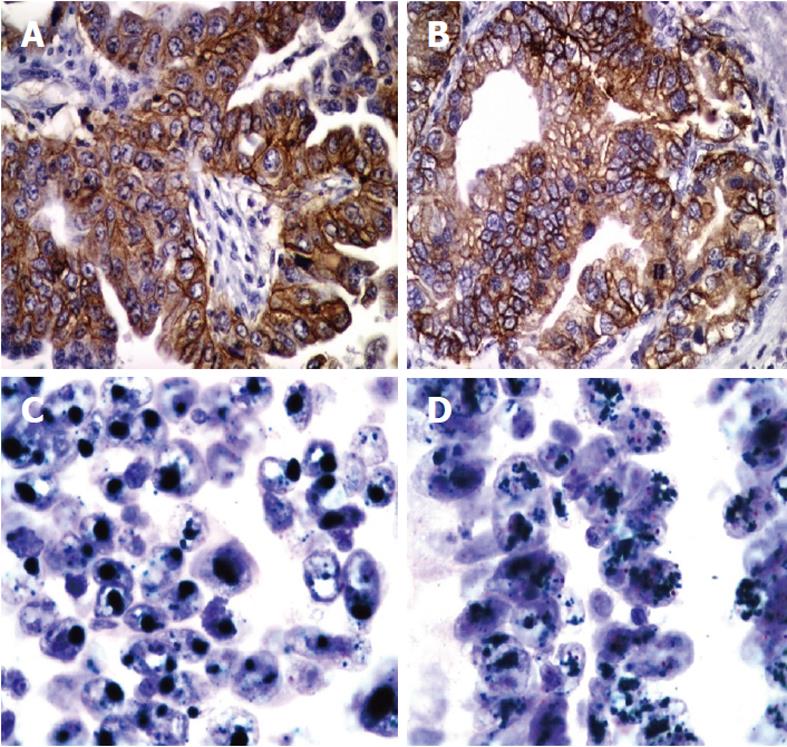
Figure 1 Examples of human epidermal growth factor receptor-2 immunohistochemical expression and amplification in primary and metastatic gastric cancer.
Immunohistochemistry shows strong membranous staining of human epidermal growth factor receptor-2 (HER2) in intestinal type gastric cancer (A) and (B) (× 400). Chromogenic in situ hybridization assay shows amplification of HER2 in primary gastric cancer (C) and the corresponding lymph node metastasis (D). Clustered green signals represent the amplified HER2 gene, while red signals represent centromere 17. Cell nuclei are counterstained with hematoxylin (× 1000).
Figure 2 Kaplan-Meier curve for disease-specific survival and human epidermal growth factor receptor-2 amplification in gastric carcinomas.
HER2: Human epidermal growth factor receptor-2.
- Citation: Tsapralis D, Panayiotides I, Peros G, Liakakos T, Karamitopoulou E. Human epidermal growth factor receptor-2 gene amplification in gastric cancer using tissue microarray technology. World J Gastroenterol 2012; 18(2): 150-155
- URL: https://www.wjgnet.com/1007-9327/full/v18/i2/150.htm
- DOI: https://dx.doi.org/10.3748/wjg.v18.i2.150


