Copyright
©2012 Baishideng Publishing Group Co.
World J Gastroenterol. Apr 21, 2012; 18(15): 1822-1826
Published online Apr 21, 2012. doi: 10.3748/wjg.v18.i15.1822
Published online Apr 21, 2012. doi: 10.3748/wjg.v18.i15.1822
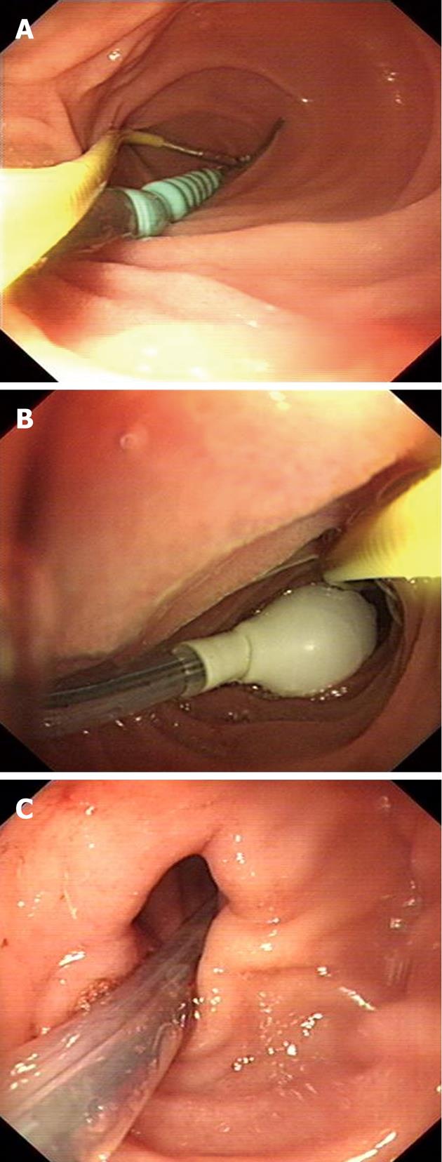
Figure 1 Endoscopic progress of a long-tube insertion.
A: The guidewire was grasped with biopsy forceps and the scope and tube were passed through the pylorus to reach the duodenojejunal flexure; B: The anterior balloon was inflated to engage the wall of the bowel; C: The guidewire was released and the scope was withdrawn while maintaining the long tube in the small bowel.
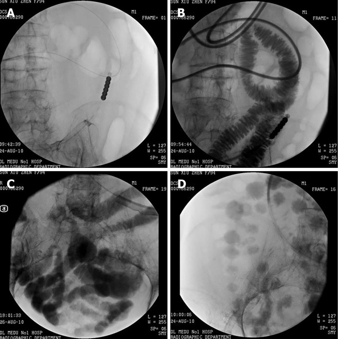
Figure 2 Abdominal flat plate images after long tube insertion.
A: Location of the long tube; B: Jejunum after ingestion of contrast medium through the long tube; C: Ileum; D: Colon, showing complete relief of the small bowel obstruction after insertion of the long tube.
- Citation: Guo SB, Duan ZJ. Decompression of the small bowel by endoscopic long-tube placement. World J Gastroenterol 2012; 18(15): 1822-1826
- URL: https://www.wjgnet.com/1007-9327/full/v18/i15/1822.htm
- DOI: https://dx.doi.org/10.3748/wjg.v18.i15.1822


