Copyright
©2012 Baishideng Publishing Group Co.
World J Gastroenterol. Mar 21, 2012; 18(11): 1208-1215
Published online Mar 21, 2012. doi: 10.3748/wjg.v18.i11.1208
Published online Mar 21, 2012. doi: 10.3748/wjg.v18.i11.1208
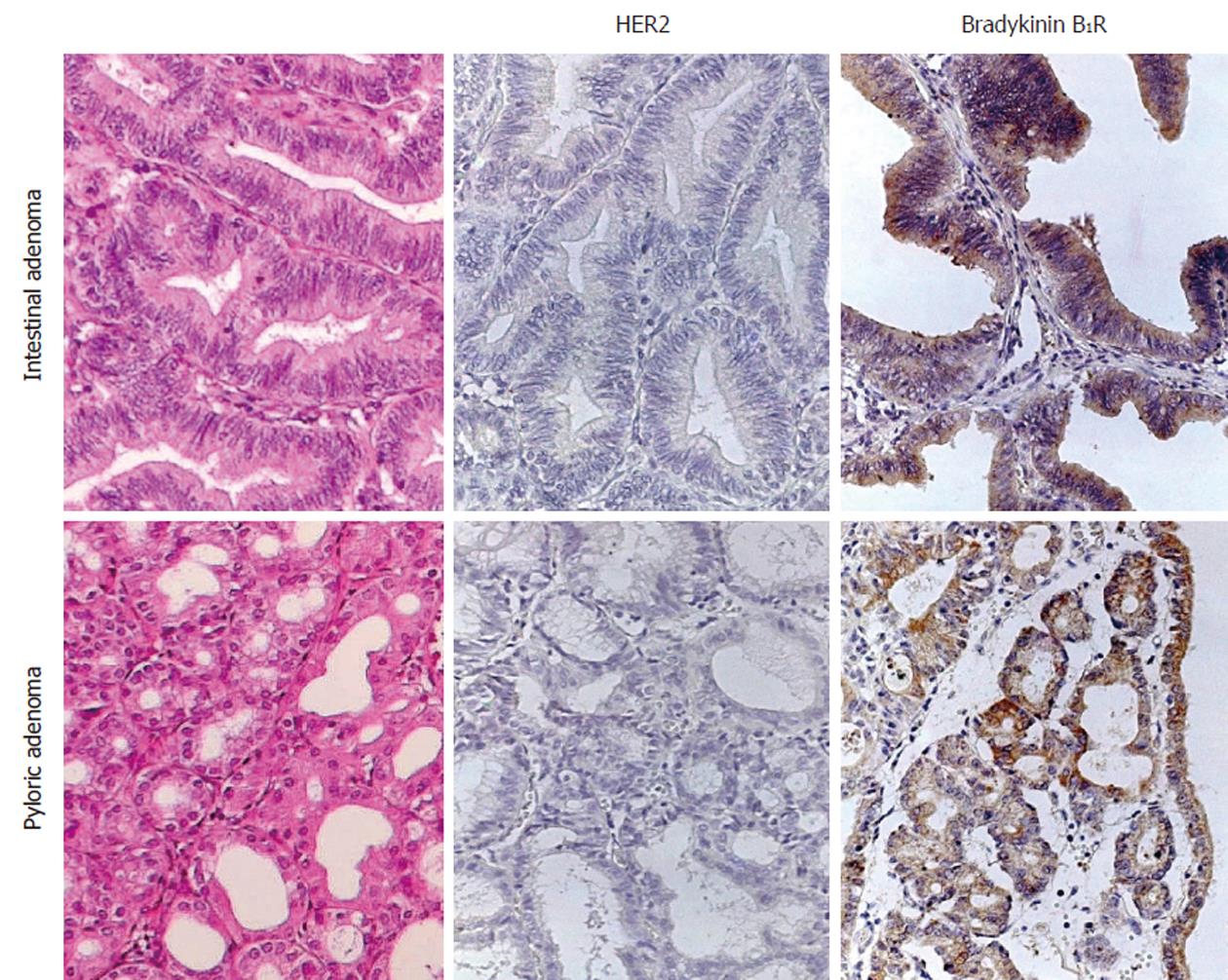
Figure 1 Expression of immunoreactive HER2 and bradykinin B1 receptor in pyloric- and intestinal-type adenomas.
Tissue sections were incubated with each antibody and then the biotin/streptavidin-peroxidase technique was followed. B1R: B1 receptor.
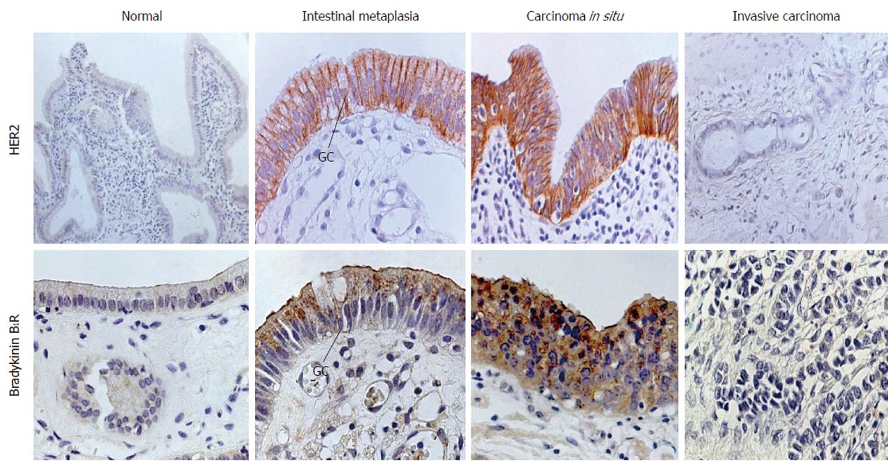
Figure 2 Immunoreactive HER2 and bradykinin B1 receptor receptors in normal gallbladder, invasive carcinoma and in epithelia with intestinal metaplasia and carcinoma in situ.
Biotin/streptavidin-peroxidase technique. B1R: B1 receptor; GC: Goblet cell.
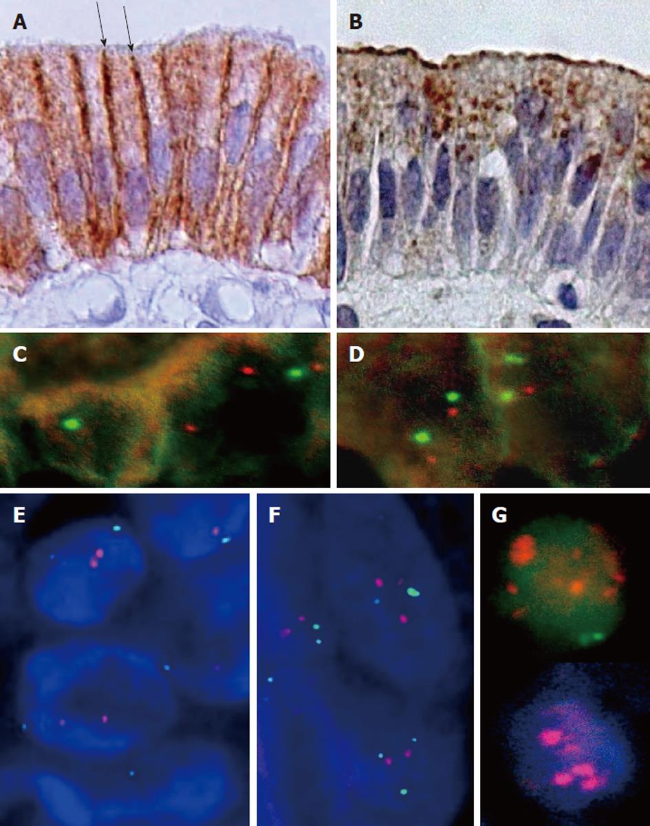
Figure 3 High magnification of gallbladder metaplastic epithelium showing immunoreactivity for HER2 (A) and bradykinin B1 receptor receptor (B).
Arrows show the limit between apical and basolateral cell membrane domains. C-G: Fluorescence in situ hybridization for HER2. Tissue sections were hybridized with a mixture of HER2-Texas Red and cen-17 labeled with fluorescein isothiocyanate. C: Normal gallbladder epithelium; D: Epithelium with intestinal metaplasia; E: Carcinoma in situ; F: Invasive carcinoma; G: Positive control corresponding to a breast cancer sample classified as HER2 + 3.
- Citation: Toledo C, Matus CE, Barraza X, Arroyo P, Ehrenfeld P, Figueroa CD, Bhoola KD, del Pozo M, Poblete MT. Expression of HER2 and bradykinin B1 receptors in precursor lesions of gallbladder carcinoma. World J Gastroenterol 2012; 18(11): 1208-1215
- URL: https://www.wjgnet.com/1007-9327/full/v18/i11/1208.htm
- DOI: https://dx.doi.org/10.3748/wjg.v18.i11.1208


