Copyright
©2012 Baishideng Publishing Group Co.
World J Gastroenterol. Mar 14, 2012; 18(10): 1123-1129
Published online Mar 14, 2012. doi: 10.3748/wjg.v18.i10.1123
Published online Mar 14, 2012. doi: 10.3748/wjg.v18.i10.1123
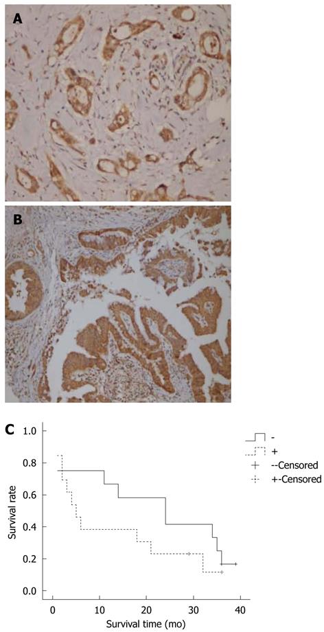
Figure 1 Interleukin-8 and CXCR1 expression in pancreatic cancer.
A: Interleukin-8 (IL-8) expression in pancreatic cancer (× 200); B: CXCR1 expression in pancreatic cancer (× 200); The positive signals of IL-8 and CXCR1 were mainly located in the cytoplasm of cancer cells. For IL-8, focal positivity was found in the fibroblasts; C: Survival analysis of pancreatic cancer patients. “Censored” means patients were still alive till the end of follow-up study. The patients without IL-8 expression survived longer than those IL-8 positive.
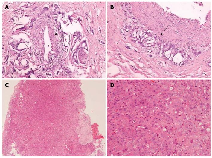
Figure 2 Histology of human pancreatic cancer tissue and transplanted tumor in SCID mice.
A: Both vascular (arrow) and neural invasion (cross) were seen in the original human pancreatic ductal adenocarcinoma (PDC) (× 100); B: Cancer cells were infiltrating the medium-sized vessel in human PDC tissues (× 100) (arrow). Desmoplastic response was obvious in the human PDC tissues; C, D: Transplanted cancer tissue in SCID mice in low power (C, × 40) and in high power (D, × 200). Cancer cells were almost clustered and poorly differentiated. In some areas, primitive gland formation could be seen (arrow). Cells grew into sheet with very less stromal reaction than the original human tissues.
- Citation: Chen Y, Shi M, Yu GZ, Qin XR, Jin G, Chen P, Zhu MH. Interleukin-8, a promising predictor for prognosis of pancreatic cancer. World J Gastroenterol 2012; 18(10): 1123-1129
- URL: https://www.wjgnet.com/1007-9327/full/v18/i10/1123.htm
- DOI: https://dx.doi.org/10.3748/wjg.v18.i10.1123


