Copyright
©2012 Baishideng Publishing Group Co.
World J Gastroenterol. Jan 7, 2012; 18(1): 79-83
Published online Jan 7, 2012. doi: 10.3748/wjg.v18.i1.79
Published online Jan 7, 2012. doi: 10.3748/wjg.v18.i1.79
Figure 1 Effects of 4-phenyl butyric acid on the proliferation of gastric carcinoma MGC-803 cells (A) and SGC-7901 cells (B).
Cells were incubated with 4-phenyl butyric acid at various concentrations for 24, 48, 72, and 96 h. The proliferation of cells was determined by the MTT assay. aP < 0.05, bP < 0.01 vs control. The relative inhibition rate was calculated as a percentage, as follows: (1-Aexperiment/Acontrol) × 100%.
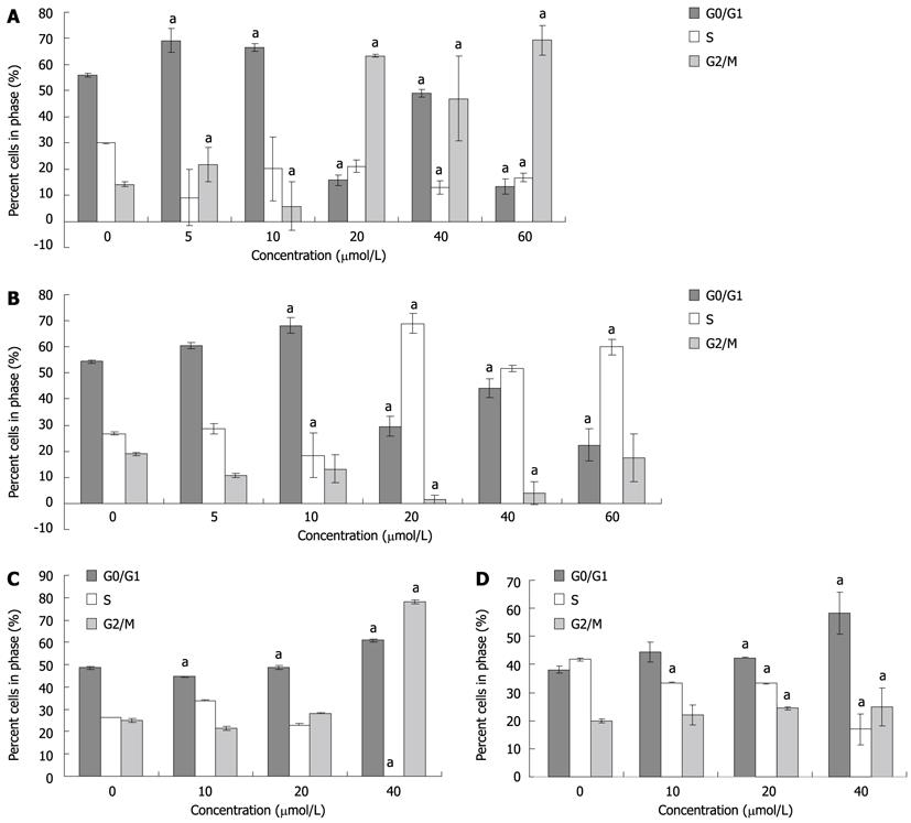
Figure 2 Effects of 4-phenyl butyric acid on cell cycle distribution of gastric carcinoma cells.
The cell cycle was measured by propidium iodide staining and fluorescence-activated cell sorting analysis. aP < 0.05 vs control. MGC-803 cells (A) and SGC-7901 cells (B) were treated for 48 h; and MGC-803 cells (C) and SGC-7901 cells (D) were treated for 24 h.
- Citation: Li LZ, Deng HX, Lou WZ, Sun XY, Song MW, Tao J, Xiao BX, Guo JM. Growth inhibitory effect of 4-phenyl butyric acid on human gastric cancer cells is associated with cell cycle arrest. World J Gastroenterol 2012; 18(1): 79-83
- URL: https://www.wjgnet.com/1007-9327/full/v18/i1/79.htm
- DOI: https://dx.doi.org/10.3748/wjg.v18.i1.79


