Copyright
©2011 Baishideng Publishing Group Co.
World J Gastroenterol. Feb 28, 2011; 17(8): 1051-1057
Published online Feb 28, 2011. doi: 10.3748/wjg.v17.i8.1051
Published online Feb 28, 2011. doi: 10.3748/wjg.v17.i8.1051
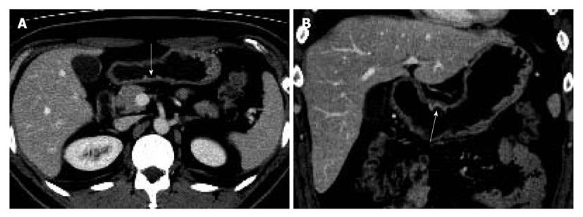
Figure 1 Early gastric cancer in a 45-year-old man.
The size of tumor was 3.8 cm at the longest diameter and the tumor extended to submucosal layer. The transverse (A) and coronal multiplanar (B) images show an ulcerative, thickened wall with enhancement in posterior wall of gastric antrum (arrows). It was graded as 4 at lower 1/3 segment of stomach on both blinded and unblinded analysis by both reviewers.
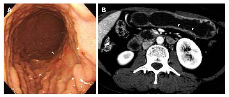
Figure 2 Early gastric cancer in a 45-year-old woman.
The size of tumor was 9.5 cm at the longest diameter and the tumor extended to submucosal layer. A: The gastroscopy shows a large ill-defined lesion with abnormal convergence of gastric folds in posterior wall of gastric mid body (arrowheads); B: The transverse computed tomography scan shows equivocal enhancement and thickening (arrowheads) in posterior wall of gastric mid body. Although one reviewer indicated this lesion on both blinded and unblinded analysis, the other did not find this lesion even on unblinded analysis.
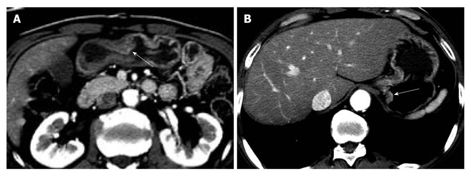
Figure 3 Early gastric cancer in a 62-year-old man.
The size of tumor was 2.2 cm at the longest diameter and the tumor extended to submucosal layer. A: The transverse computed tomography scan shows thickened wall with enhancement in greater curvature of gastric antrum (arrow), which was true lesion based on gastroscopy and pathologic examination; B: Reviewer 1 indicated a lesion (arrow) as a cancer focus because thickened wall was suspicious on blinded analysis. However, on unblinded analysis, focal lesion (arrow) in greater curvature of gastric antrum in lower gastric 1/3 segment was indicated correctly.
- Citation: Park KJ, Lee MW, Koo JH, Park Y, Kim H, Choi D, Lee SJ. Detection of early gastric cancer using hydro-stomach CT: Blinded vs unblinded analysis. World J Gastroenterol 2011; 17(8): 1051-1057
- URL: https://www.wjgnet.com/1007-9327/full/v17/i8/1051.htm
- DOI: https://dx.doi.org/10.3748/wjg.v17.i8.1051


