Copyright
©2011 Baishideng Publishing Group Co.
World J Gastroenterol. Jan 21, 2011; 17(3): 366-371
Published online Jan 21, 2011. doi: 10.3748/wjg.v17.i3.366
Published online Jan 21, 2011. doi: 10.3748/wjg.v17.i3.366
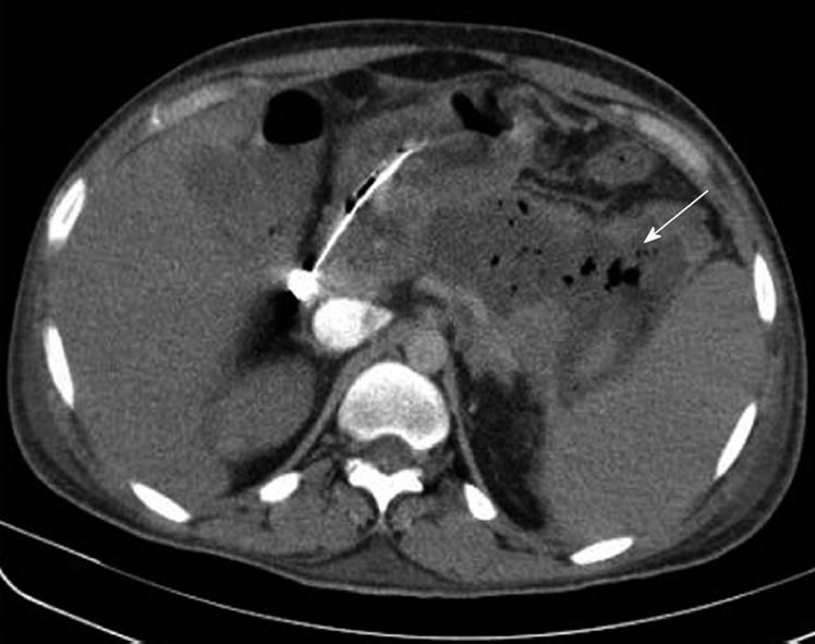
Figure 1 Contrast-enhanced computed tomography of the abdomen showing a large hypodense collection with air pockets in the location of the pancreatic body and tail (white arrow) indicative of an infected pancreatic necrosis.
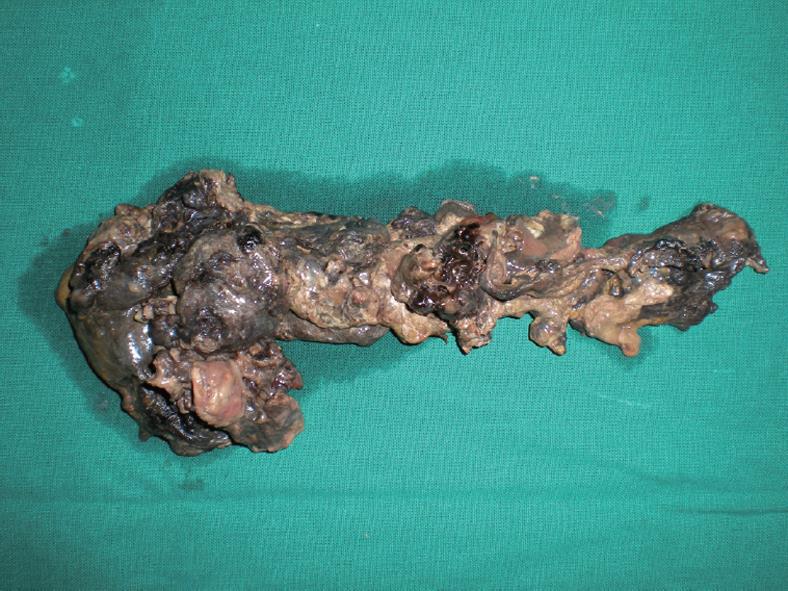
Figure 2 Post-operative photograph demonstrating a complete necrotic pancreas.
- Citation: Doctor N, Philip S, Gandhi V, Hussain M, Barreto SG. Analysis of the delayed approach to the management of infected pancreatic necrosis. World J Gastroenterol 2011; 17(3): 366-371
- URL: https://www.wjgnet.com/1007-9327/full/v17/i3/366.htm
- DOI: https://dx.doi.org/10.3748/wjg.v17.i3.366


