Copyright
©2011 Baishideng Publishing Group Co.
World J Gastroenterol. Jan 21, 2011; 17(3): 283-289
Published online Jan 21, 2011. doi: 10.3748/wjg.v17.i3.283
Published online Jan 21, 2011. doi: 10.3748/wjg.v17.i3.283
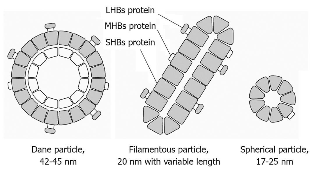
Figure 1 Schematic model of hepatitis B surface antigen structure.
Three forms of hepatitis B surface (HBs) antigen (Dane particle, filamentous particle, and spherical particle) are visualized in serum by electron microscopy. These are composed of small, middle, and large hepatitis B surface proteins. LHBs: Large HBs proteins; MHBs: Middle HBs proteins; SHBs: Small HBs proteins.
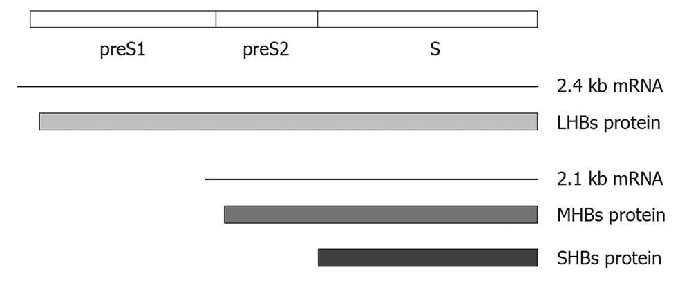
Figure 2 Schematic presentation of the S/preS1/preS2 gene, RNA transcripts, and translational products.
Opening reading frame S has three internal AUG codons. Transcription to produce the 2.1 kb and 2.4 kb mRNAs first occurs after translation into small hepatitis B surface proteins (SHBs), middle hepatitis B surface proteins (MHBs), and large hepatitis B surface proteins (LHBs) ensues with different promoters.
- Citation: Lee JM, Ahn SH. Quantification of HBsAg: Basic virology for clinical practice. World J Gastroenterol 2011; 17(3): 283-289
- URL: https://www.wjgnet.com/1007-9327/full/v17/i3/283.htm
- DOI: https://dx.doi.org/10.3748/wjg.v17.i3.283


