Copyright
©2011 Baishideng Publishing Group Co.
World J Gastroenterol. Jan 14, 2011; 17(2): 267-270
Published online Jan 14, 2011. doi: 10.3748/wjg.v17.i2.267
Published online Jan 14, 2011. doi: 10.3748/wjg.v17.i2.267
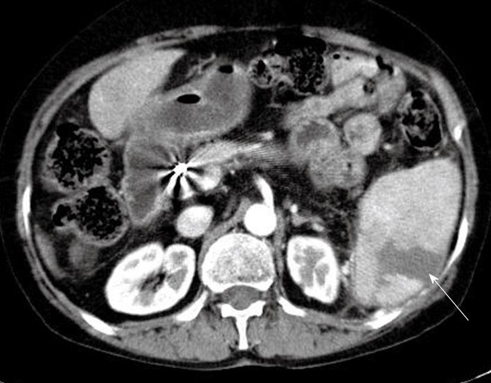
Figure 1 Contrast-enhanced abdominal computed tomography.
A wedge-shaped hypodense lesion in the spleen is consistent with splenic infarction (arrow). Hepatic artery catheter is seen in hepatic artery as artifact.
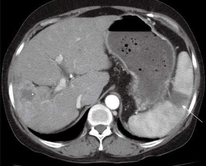
Figure 2 Follow up abdominal computed tomography (1 mo).
New hypodense lesion has developed in upper level of the spleen (arrow).
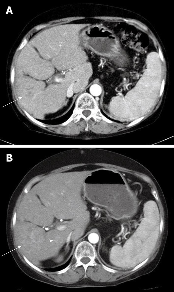
Figure 3 Contrast-enhanced computed tomography scan in arterial phase at the time of diagnosis of initial splenic infarction (A) and 2 mo later (B).
A: When splenic infarction was diagnosed, an approximately 4 cm x 4 cm sized hepatocellular carcinoma (HCC) (arrow) was shown in S6; B: Two months later, the size of HCC was enlarged to 5 cm × 5 cm (arrow) and an intrahepatic metastatic nodule was also seen (arrowhead) beside main mass.
- Citation: Kim SO, Han SY, Baek YH, Lee SW, Han JS, Kim BG, Cho JH, Nam KJ. Splenic infarction associated with sorafenib use in a hepatocellular carcinoma patient. World J Gastroenterol 2011; 17(2): 267-270
- URL: https://www.wjgnet.com/1007-9327/full/v17/i2/267.htm
- DOI: https://dx.doi.org/10.3748/wjg.v17.i2.267


