Copyright
©2011 Baishideng Publishing Group Co.
World J Gastroenterol. Jan 14, 2011; 17(2): 219-225
Published online Jan 14, 2011. doi: 10.3748/wjg.v17.i2.219
Published online Jan 14, 2011. doi: 10.3748/wjg.v17.i2.219
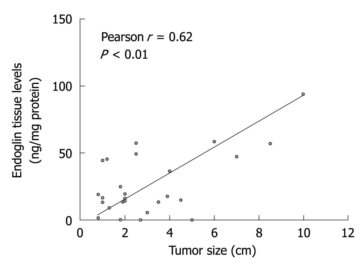
Figure 1 Orthogonal regression analysis of endoglin tissue levels and tumor size (n = 26) in 17 patients.
(In one patient, information about tumor size was missing, so this patient was not included in this analysis). Increasing endoglin levels in tumors were significantly correlated with greater tumor size.
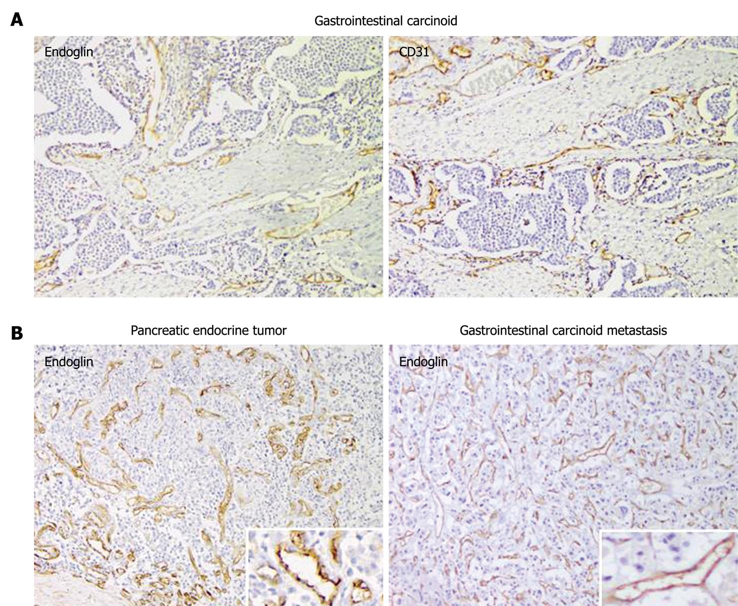
Figure 2 Immunostaining of endoglin and CD31 on peritumoral and intratumoral vessels in gastroenteropancreatic neuroendocrine tumors.
A: Endoglin staining was limited to angiogenic vessels, whereas CD31 stained both old and new blood vessels in tumor tissue. Magnification 100 ×; B: Representative endoglin staining in a pancreatic neuroendocrine tumor and a gastrointestinal carcinoid metastasis (small bowel mesentery). Magnification 100 ×. Inserts show a higher magnification at 200 ×.
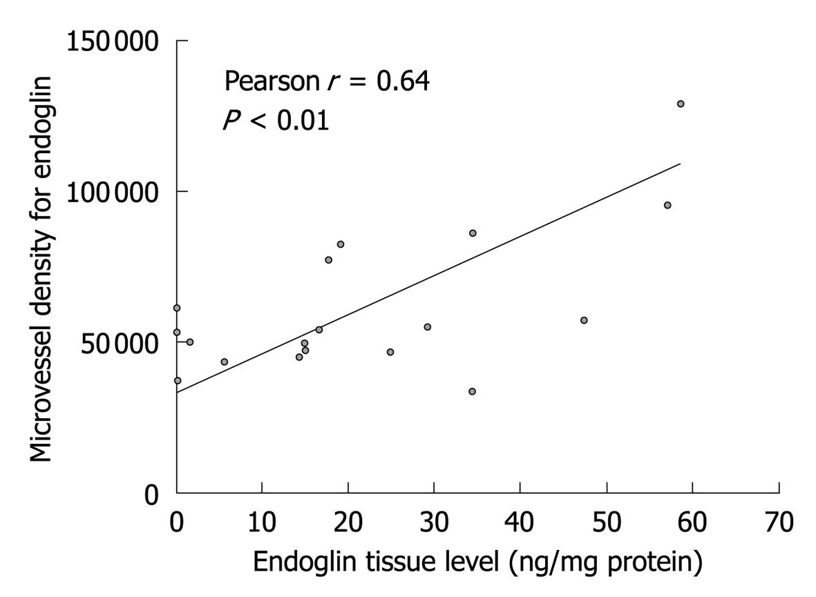
Figure 3 Correlation analysis of the endoglin microvessel density and endoglin tissue levels in tumors (n = 17).
For one patient in whom endoglin tissue levels were assessed, no paraffin slides for microvessel density (MVD) determination were available. Endoglin MVD was significantly correlated with tumor levels of endoglin.
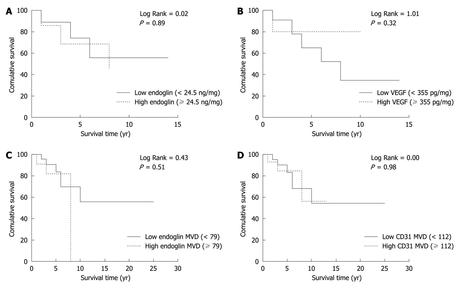
Figure 4 Kaplan-Meier survival analysis for endoglin tumor levels (A), vascular endothelial growth factor tumor levels (B), endoglin microvessel density (C) and CD31 microvessel density (D).
Patients were divided into two groups based on mean tumor levels (A and B) or mean microvessel density (MVD) scores (C and D). None of the parameters showed a significant correlation with patient survival. VEGF: Vascular endothelial growth factor.
- Citation: Kuiper P, Hawinkels LJ, Jonge-Muller ES, Biemond I, Lamers CB, Verspaget HW. Angiogenic markers endoglin and vascular endothelial growth factor in gastroenteropancreatic neuroendocrine tumors. World J Gastroenterol 2011; 17(2): 219-225
- URL: https://www.wjgnet.com/1007-9327/full/v17/i2/219.htm
- DOI: https://dx.doi.org/10.3748/wjg.v17.i2.219


