Copyright
©2011 Baishideng Publishing Group Co.
World J Gastroenterol. May 21, 2011; 17(19): 2365-2371
Published online May 21, 2011. doi: 10.3748/wjg.v17.i19.2365
Published online May 21, 2011. doi: 10.3748/wjg.v17.i19.2365
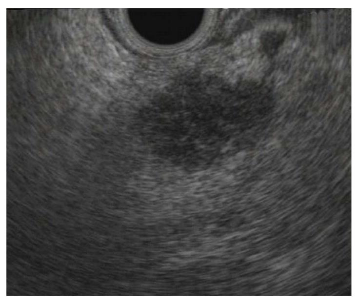
Figure 1 Radial endoscopic ultrasound image of a small (12 mm) early pancreatic cancer, seen as an irregular hypoechoic mass lesion in the pancreatic head.
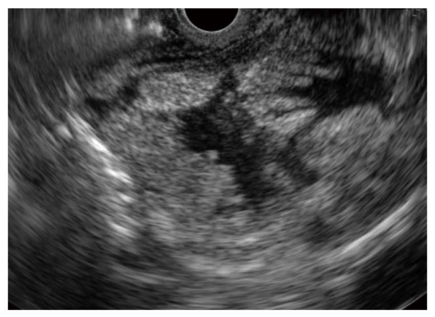
Figure 2 Endoscopic ultrasound image of 17 mm × 7 mm main-duct type intraductal papillary mucinous neoplasm.
The duct is markedly dilated and villous papillary projections can be seen arising from the duct wall. Pathology after surgical resection showed high-grade dysplasia.
- Citation: Stoita A, Penman ID, Williams DB. Review of screening for pancreatic cancer in high risk individuals. World J Gastroenterol 2011; 17(19): 2365-2371
- URL: https://www.wjgnet.com/1007-9327/full/v17/i19/2365.htm
- DOI: https://dx.doi.org/10.3748/wjg.v17.i19.2365


