Copyright
©2011 Baishideng Publishing Group Co.
World J Gastroenterol. May 14, 2011; 17(18): 2332-2337
Published online May 14, 2011. doi: 10.3748/wjg.v17.i18.2332
Published online May 14, 2011. doi: 10.3748/wjg.v17.i18.2332
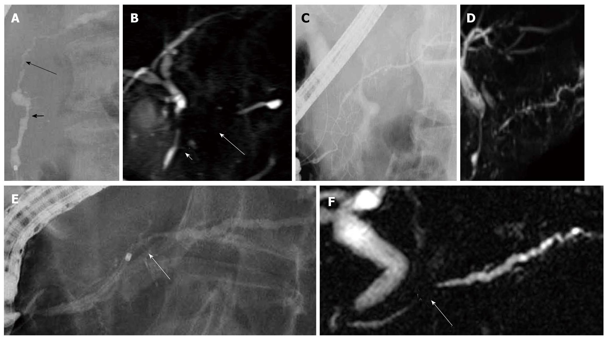
Figure 1 Pancreatography of autoimmune pancreatitis.
A: Endoscopic retrograde cholangiopancreatography finding of autoimmune pancreatitis showing skipped lesions of the main pancreatic duct (short and long arrows); B: On magnetic resonance cholangiopancreatography, skipped lesions (short and long arrows) on endoscopic retrograde cholangiopancreatography were not visualized; C: Endoscopic retrograde cholangiopancreatography finding of autoimmune pancreatitis showing side branch derivation from the narrowed portion of the main pancreatic duct; D: Recent magnetic resonance cholangiopancreatography could show diffuse narrowing of the main pancreatic duct fairly well; E: Endoscopic retrograde cholangiopancreatography finding of autoimmune pancreatitis showing a short narrowed main pancreatic duct (arrow) with upstream dilatation less than 5 mm; F: On magnetic resonance cholangiopancreatography, the narrowed portion (arrow) was not visualized.
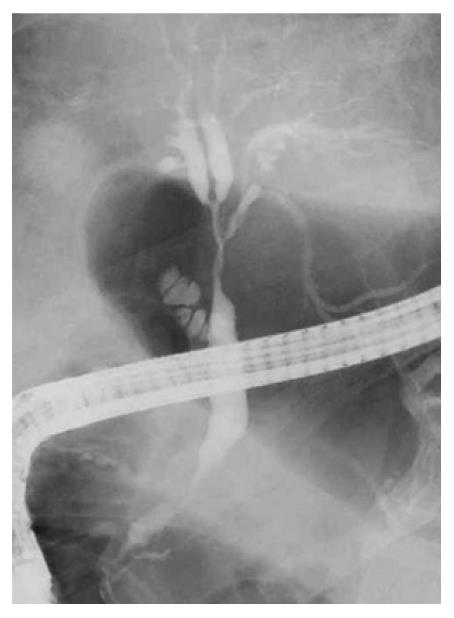
Figure 2 Endoscopic retrograde cholangiopancreatography finding of autoimmune pancreatitis showing stenosis of the hilar and intrahepatic bile duct.
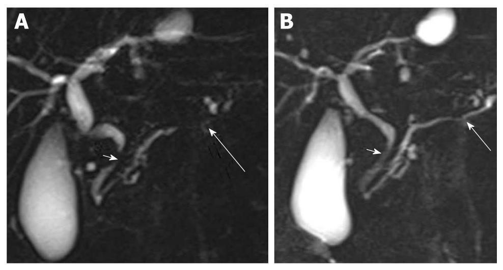
Figure 3 Magnetic resonance cholangiopancreatography finding of autoimmune pancreatitis.
Stenosis of the lower bile duct (short arrow) and the narrowing of the main pancreatic duct (long arrow) (A) improved after steroid therapy (B).
- Citation: Takuma K, Kamisawa T, Tabata T, Inaba Y, Egawa N, Igarashi Y. Utility of pancreatography for diagnosing autoimmune pancreatitis. World J Gastroenterol 2011; 17(18): 2332-2337
- URL: https://www.wjgnet.com/1007-9327/full/v17/i18/2332.htm
- DOI: https://dx.doi.org/10.3748/wjg.v17.i18.2332


