Copyright
©2011 Baishideng Publishing Group Co.
World J Gastroenterol. May 14, 2011; 17(18): 2315-2325
Published online May 14, 2011. doi: 10.3748/wjg.v17.i18.2315
Published online May 14, 2011. doi: 10.3748/wjg.v17.i18.2315
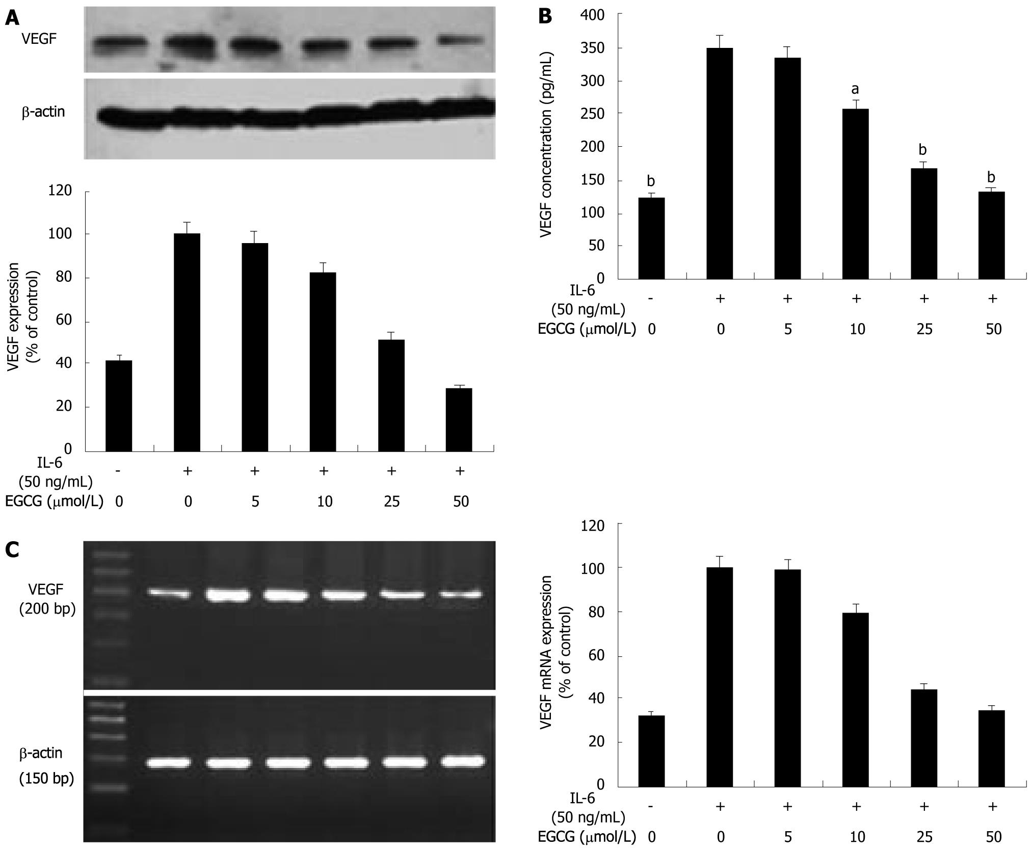
Figure 1 (-)-Epigallocatechin-3-gallate inhibits vascular endothelial growth factor production induced by interleukin-6 in human gastric cancer cells.
A: When treated with (-)-Epigallocatechin-3-gallate (EGCG), vascular endothelial growth factor (VEGF) expression was dose-dependently decreased; B: Enzyme-linked immunosorbent assay showed that interleukin-6 (IL-6) induced VEGF secretion in human gastric cancer (AGS) cells and a 2.8-fold increase was observed. EGCG treatment dose-dependently reduced VEGF protein level in the conditioned medium. C: IL-6 also induced VEGF mRNA expression and a 3.1-fold increase in VEGF mRNA expression was observed. When treated with EGCG, VEGF mRNA expression was dose-dependently decreased. Values are expressed as percent of control (means ± SE, n = 3, aP < 0.05, bP < 0.01).
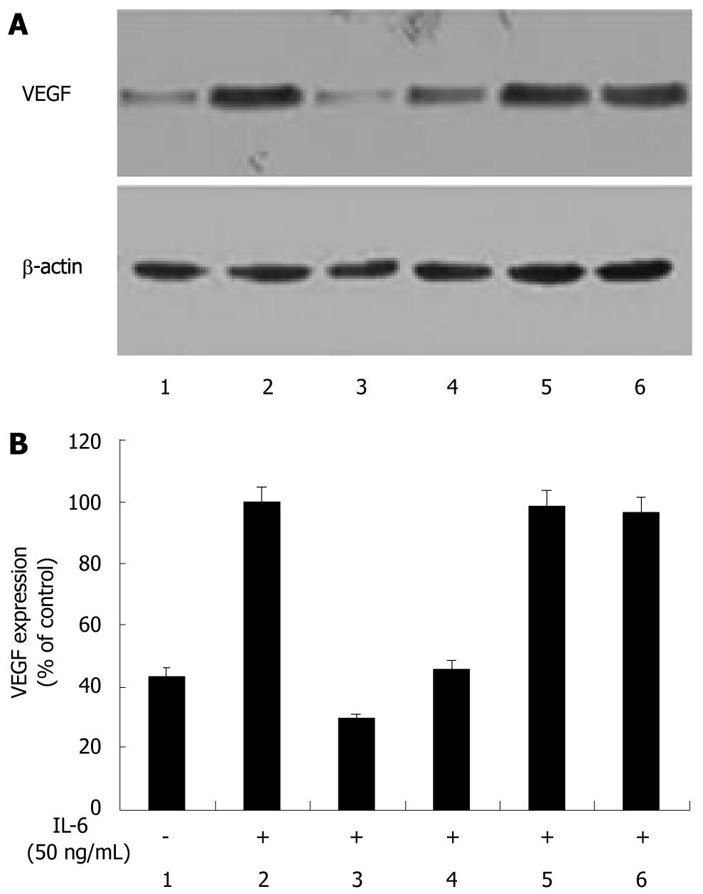
Figure 2 (-)-Epigallocatechin-3-gallate inhibits interleukin-6-induced vascular endothelial growth factor expression in human gastric cancer cells via JAK/STAT pathway.
Human gastric cancer (AGS) cells were stimulated with interleukin-6 (IL-6) (50 ng/mL) for 24 h in the presence of 50 μmol (-)-epigallocatechin-3-gallate (EGCG) or signaling inhibitors. Vascular endothelial growth factor (VEGF) protein levels in tumor cell lysates were analyzed by Western blotting. IL-6 markedly increased VEGF expression in AGS cells. When treated with EGCG or AG490, VEGF expression was significantly reduced. PD98059 and LY294002 did not change IL-6-induced VEGF expression. 1: Without IL-6 stimulation; 2: Stimulated with IL-6; 3-6: Treated with 50 μmol EGCG or signaling inhibitors of 20 μmol AG490, 25 μmol PD98059 or 25 μmol LY294002.
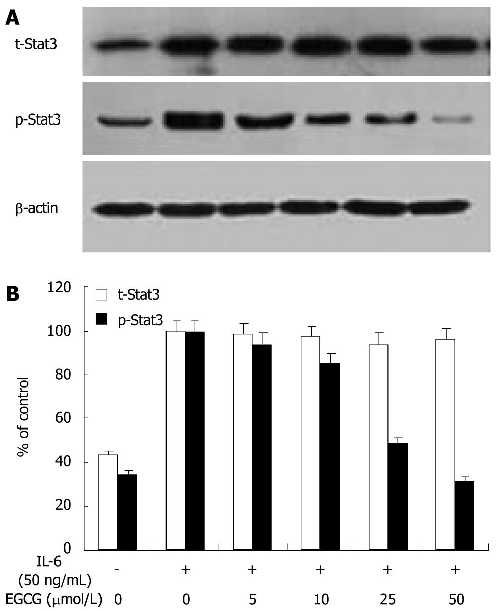
Figure 3 (-)-Epigallocatechin-3-gallate inhibits signal transducer and activator of transcription 3 activation induced by interleukin-6 in human gastric cancer cells.
Human gastric cancer (AGS) cells were stimulated with interleukin-6 (IL-6) (50 ng/mL) in the presence of (-)-epigallocatechin-3-gallate (EGCG) at concentrations indicated for 1 h. Total signal transducer and activator of transcription 3 (Stat3) and activated Stat3 were examined by Western blotting. Stat3 was constitutively activated in AGS cells. After stimulated with IL-6, total Stat3 and activated Stat3 expression were both markedly increased by 2.3 folds and 2.9 folds, respectively. When treated with EGCG, activated Stat3 expression decreased in a dose-dependant manner, but total Stat3 expression remained unchanged.
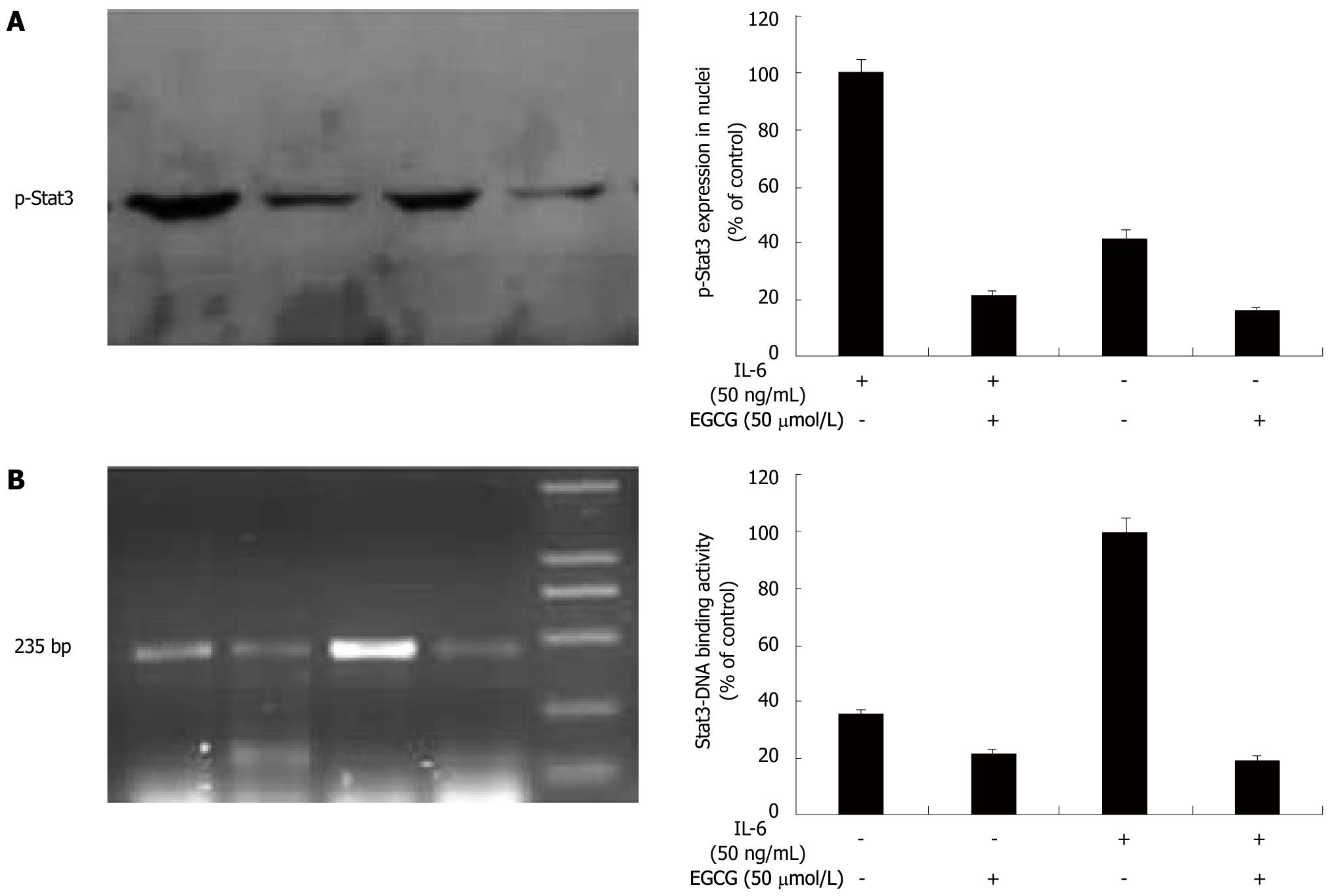
Figure 4 (-)-Epigallocatechin-3-gallate inhibits signal transducer and activator of transcription 3 nuclear translocation and DNA binding activity in human gastric cancer cells.
Signal transducer and activator of transcription 3 (Stat3) nuclear translocation was determined by Western blotting with extraction of nuclear proteins. A: After treated with 50 μmol (-)-epigallocatechin-3-gallate (EGCG) and interleukin-6 (IL-6) (50 ng/mL) for 1 h, phospho-Stat3 in the nucleus was visualized with an anti-p-Stat3 antibody; B: IL-6 apparently increased phospho-Stat3 expression in the nucleus, but EGCG treatment markedly decreased this effect. Stat3-DNA binding activity was determined by chromatin immunoprecipitation (ChIP) assay. Immunoprecipitation was conducted with Stat3 antibody followed by polymerase chain reaction (PCR) using oligonucleotide primers that yielded a 235 bp band spanning Stat3 binding site in vascular endothelial growth factor promoter. IL-6 apparently increased Stat3-DNA binding activity. When treated with EGCG, Stat3-DNA binding activity was also markedly decreased.
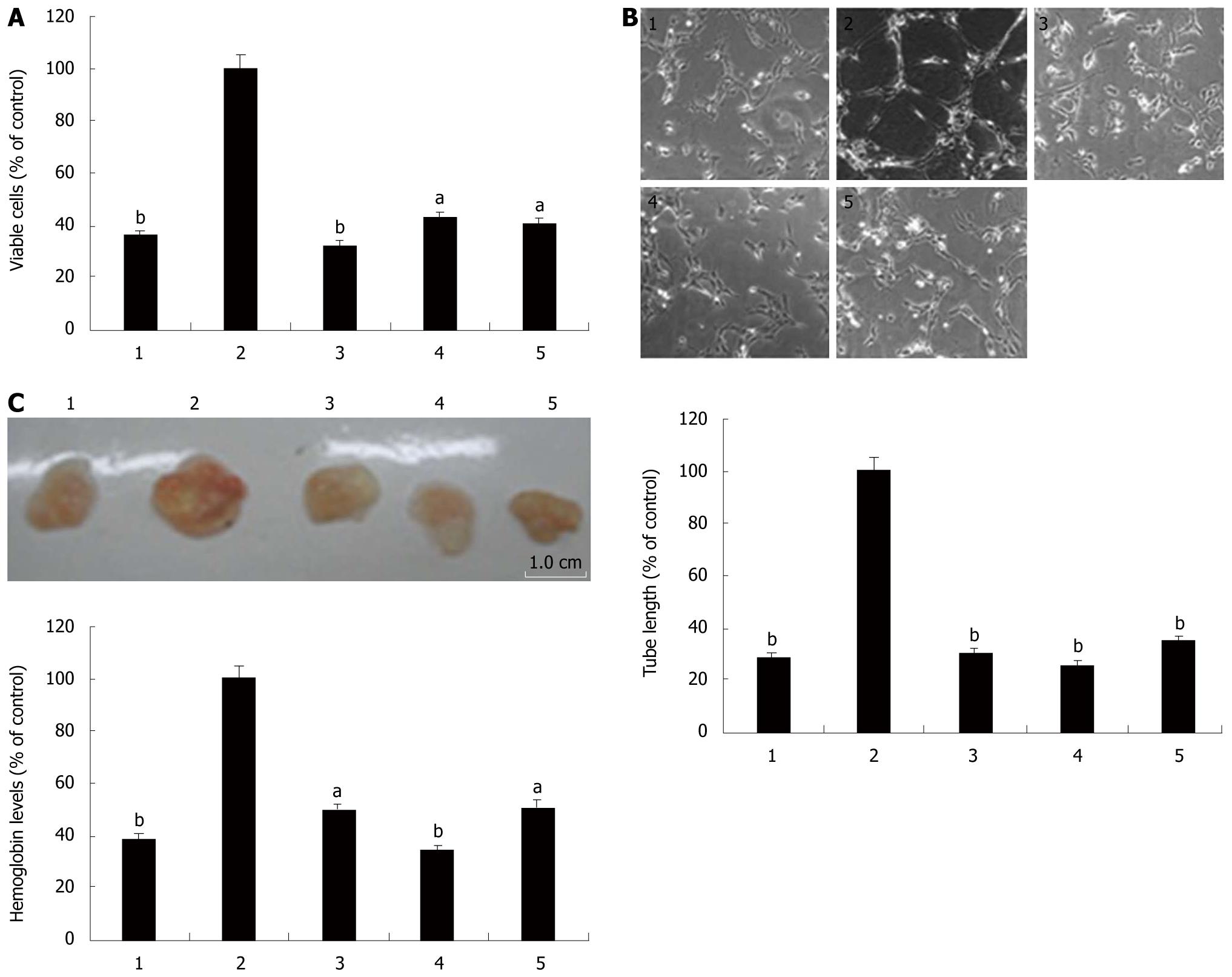
Figure 5 (-)-Epigallocatechin-3-gallate inhibits interleukin-6 induced angiogenesis in vitro and in vivo.
The in vitro angiogenesis was determined by 3-[4, 5-dimethylthiazol-2-yl]-2, 5-diphenyl tetrazoliumbromide (MTT) and tube formation assay. Human umbilical vein endothelial cells (HUVECs) were cultured with the conditioned media in the presence of vascular endothelial growth factor (VEGF) neutralizing antibody, (-)-epigallocatechin-3-gallate (EGCG) or AG490. After 48 h incubation, the viable cells were quantified by MTT assay (A), and the formed tubes were fixed, stained, photographed and analyzed (B). The in vivo angiogenesis was determined by Matrigel plug assay. Matrigel plugs containing the conditioned media with VEGF neutralizing antibody, EGCG or AG490 were subcutaneously injected into nude mice. One week later, the Matrigel plugs were harvested and examined by measuring the density of hemoglobin (C). interleukin-6 (IL-6) apparently promoted vascular endothelial cell proliferation and tube formation in vitro and vascularization of Matrigel plugs in vivo. VEGF neutralizing antibody, EGCG and AG490 all markedly decreased these effects. 1: Without IL-6 stimulation as a negative control; 2: With IL-6 stimulation as a control; 3-5: With IL-6 stimulation and VEGF neutralizing antibody, EGCG or AG490. Values are expressed as percent of control (means ± SE, n = 3, aP < 0.05, bP < 0.01).
-
Citation: Zhu BH, Chen HY, Zhan WH, Wang CY, Cai SR, Wang Z, Zhang CH, He YL. (-)-Epigallocatechin-3-gallate inhibits VEGF expression induced by IL-6
via Stat3 in gastric cancer. World J Gastroenterol 2011; 17(18): 2315-2325 - URL: https://www.wjgnet.com/1007-9327/full/v17/i18/2315.htm
- DOI: https://dx.doi.org/10.3748/wjg.v17.i18.2315


