Copyright
©2011 Baishideng Publishing Group Co.
World J Gastroenterol. Mar 14, 2011; 17(10): 1292-1303
Published online Mar 14, 2011. doi: 10.3748/wjg.v17.i10.1292
Published online Mar 14, 2011. doi: 10.3748/wjg.v17.i10.1292
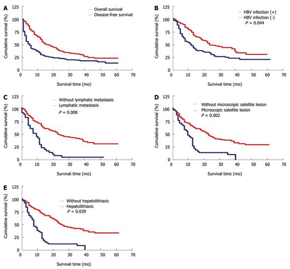
Figure 1 Overall and disease-free survival rates of patients with intrahepatic cholangiocarcinoma after surgical resection (A), higher survival rate of intrahepatic cholangiocarcinoma patients with hepatitis B virus infection than that of those without hepatitis B virus infection (B), significantly poorer survival rate of intrahepatic cholangiocarcinoma patients with lymphatic metastasis than that of those without lymphatic metastasis (C), significantly poorer survival rate of intrahepatic cholangiocarcinoma patients with microscopic satellite lesion than that of those without microscopic satellite lesion (D), and significantly poorer survival rate of intrahepatic cholangiocarcinoma patients with hepatolithiasis than that of those without hepatolithiasis (E).
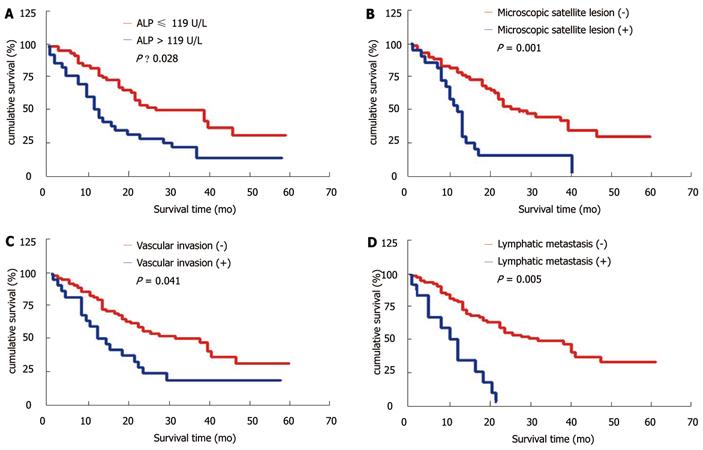
Figure 2 Adjusted survival curves according to the independent prognostic factors by multivariate analysis (Cox model) for intrahepatic cholangiocarcinoma with hepatitis B virus infection after resection.
A: Alkaline phosphatase (ALP); B: Microscopic satellite lesion; C: Vascular invasion; D: Lymphatic metastasis.
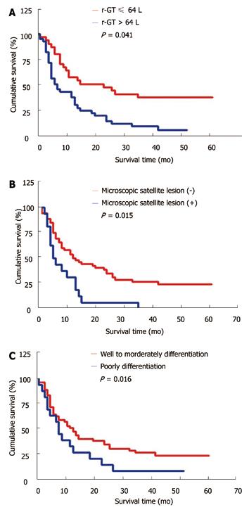
Figure 3 Adjusted survival curves according to the independent prognostic factors by multivariate analysis (Cox model) for intrahepatic cholangiocarcinoma without hepatitis B virus infection after resection.
A: R-glutamyltransferase (r-GT); B: Microscopic satellite lesion; C: Tumor differentiation.
- Citation: Zhou HB, Wang H, Li YQ, Li SX, Wang H, Zhou DX, Tu QQ, Wang Q, Zou SS, Wu MC, Hu HP. Hepatitis B virus infection: A favorable prognostic factor for intrahepatic cholangiocarcinoma after resection. World J Gastroenterol 2011; 17(10): 1292-1303
- URL: https://www.wjgnet.com/1007-9327/full/v17/i10/1292.htm
- DOI: https://dx.doi.org/10.3748/wjg.v17.i10.1292


