Copyright
©2010 Baishideng.
World J Gastroenterol. Mar 7, 2010; 16(9): 1155-1157
Published online Mar 7, 2010. doi: 10.3748/wjg.v16.i9.1155
Published online Mar 7, 2010. doi: 10.3748/wjg.v16.i9.1155
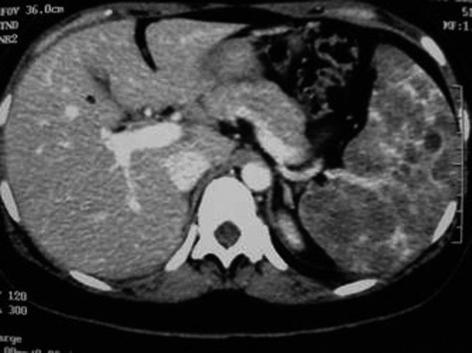
Figure 1 Total body computed tomography.
The spleen appeared increased in volume (maximum diameter 18 cm), and it was possible to see multiple low-density lesions with a nodular appearance, which replaced all the parenchyma.
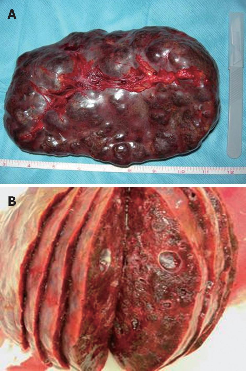
Figure 2 Surgical specimen.
A: The spleen was 18 cm on the long axis, and its macroscopic appearance was characterized by the presence of a multiple cystic mass that altered the volume and profile of the spleen; B: A section of the spleen confirmed the presence of a multiple cystic mass with a clear appearance that replaced all the parenchyma.
- Citation: Patti R, Iannitto E, Di Vita G. Splenic lymphangiomatosis showing rapid growth during lactation: A case report. World J Gastroenterol 2010; 16(9): 1155-1157
- URL: https://www.wjgnet.com/1007-9327/full/v16/i9/1155.htm
- DOI: https://dx.doi.org/10.3748/wjg.v16.i9.1155


