Copyright
©2010 Baishideng.
World J Gastroenterol. Mar 7, 2010; 16(9): 1050-1056
Published online Mar 7, 2010. doi: 10.3748/wjg.v16.i9.1050
Published online Mar 7, 2010. doi: 10.3748/wjg.v16.i9.1050
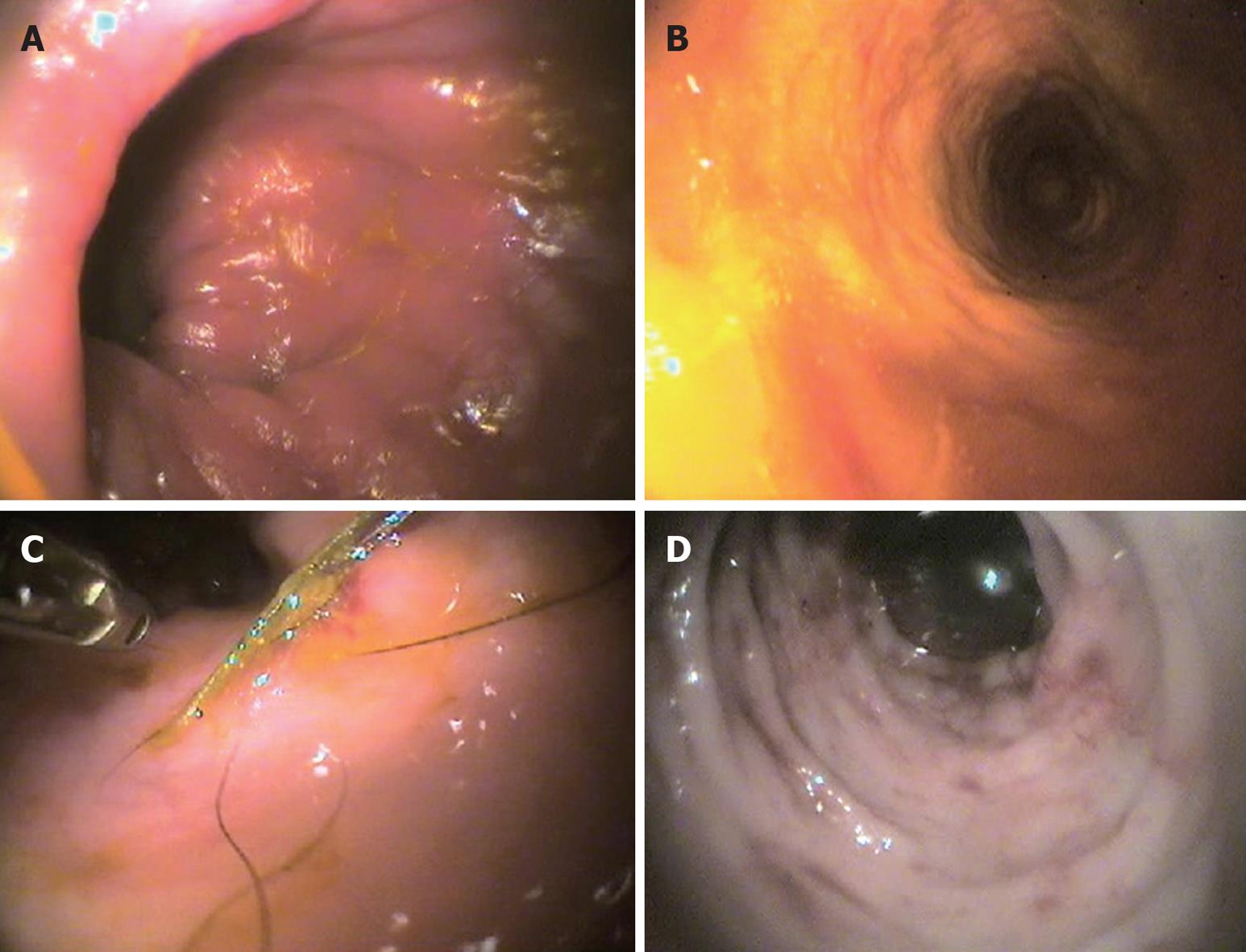
Figure 1 Representative endoscopic images in the dog.
A: Lymphocytic-plasmacytic colitis; B: Macrofollicular and diffuse interstitial lymphocytic colitis; C: Histiocytic colitis; D: Neutrophilic-eosinophilic colitis.
Figure 2 Representative histologic images in the dog (HE, bar = 50 μm).
A: Lymphocytic-plasmacytic colitis. Note the interstitial diffuse pattern of infiltrate represented by a large amount of lymphocytes mixed with plasma cells and some macrophages; B: Lymphocytic-plasmacytic colitis (follicular variant); C: Histiocytic colitis. Severe mucosal abnormalities with loss of crypts and diffuse infiltration by large macrophages (arrows) that in the insert (PAS stain) are shown as the main cells infiltrating the lamina propria; D: Eosinophilic colitis. Note the presence of a large number of eosinophils (arrows).
- Citation: Cerquetella M, Spaterna A, Laus F, Tesei B, Rossi G, Antonelli E, Villanacci V, Bassotti G. Inflammatory bowel disease in the dog: Differences and similarities with humans. World J Gastroenterol 2010; 16(9): 1050-1056
- URL: https://www.wjgnet.com/1007-9327/full/v16/i9/1050.htm
- DOI: https://dx.doi.org/10.3748/wjg.v16.i9.1050


