Copyright
©2010 Baishideng Publishing Group Co.
World J Gastroenterol. Dec 21, 2010; 16(47): 6016-6019
Published online Dec 21, 2010. doi: 10.3748/wjg.v16.i47.6016
Published online Dec 21, 2010. doi: 10.3748/wjg.v16.i47.6016
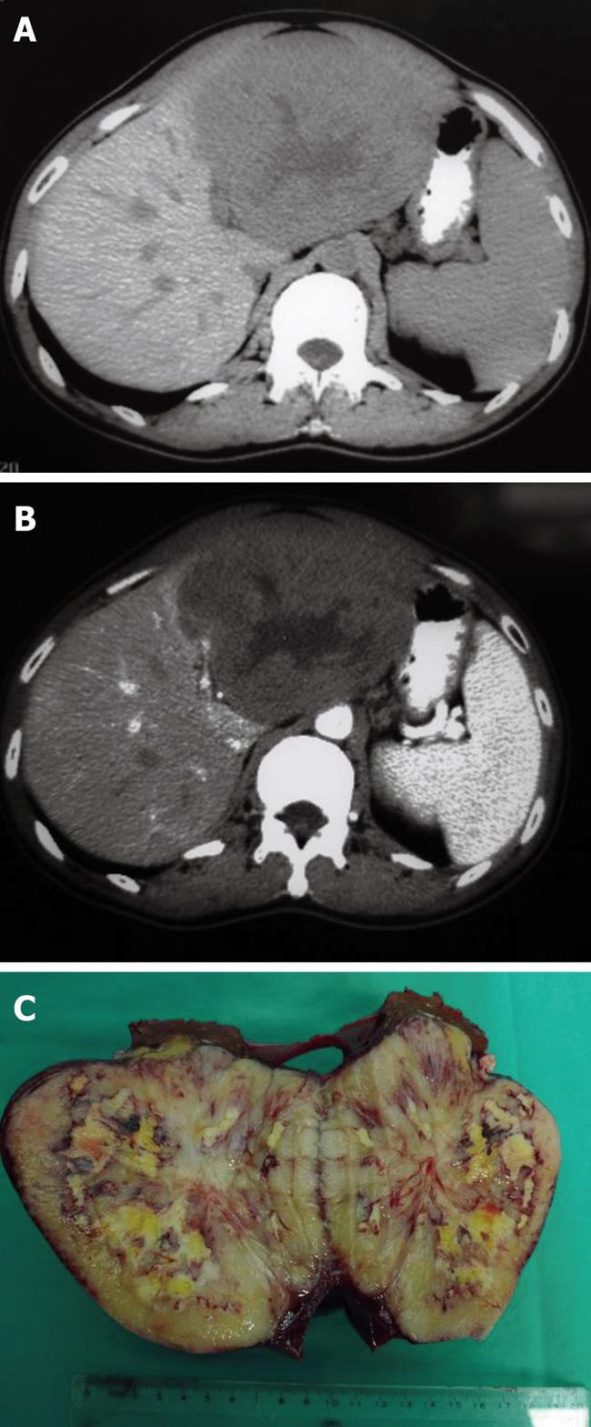
Figure 1 The preoperative imaging and surgical specimen in primary hepatic lymphoma.
A: By computed tomography, primary hepatic lymphoma (PHL) appeared as a solitary hypoattenuating lesion with a central area of low intensity; B: PHL lesion was slightly enhanced following the administration of intravenous contrast agent; C: Pathological features revealed that the PHL lesion was an encapsulated, hypovascular tumor with central necrosis.
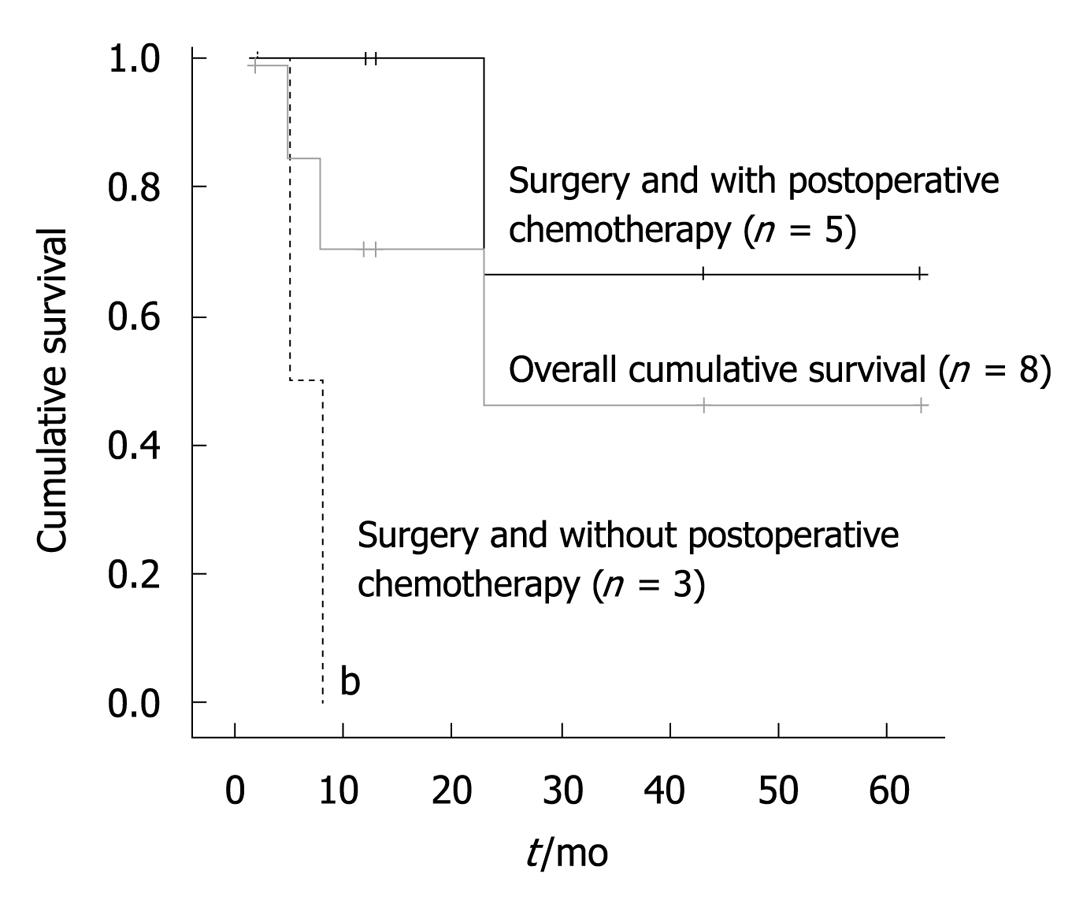
Figure 2 Survival among patients undergoing hepatic resection for primary hepatic lymphoma, with or without postoperative chemotherapy, and the overall cumulative survival of all patients (except for one who died perioperatively).
bP < 0.01 vs hepatic resection for primary hepatic lymphoma with postoperative chemotherapy (log rank test).
- Citation: Yang XW, Tan WF, Yu WL, Shi S, Wang Y, Zhang YL, Zhang YJ, Wu MC. Diagnosis and surgical treatment of primary hepatic lymphoma. World J Gastroenterol 2010; 16(47): 6016-6019
- URL: https://www.wjgnet.com/1007-9327/full/v16/i47/6016.htm
- DOI: https://dx.doi.org/10.3748/wjg.v16.i47.6016


