Copyright
©2010 Baishideng Publishing Group Co.
World J Gastroenterol. Dec 7, 2010; 16(45): 5739-5745
Published online Dec 7, 2010. doi: 10.3748/wjg.v16.i45.5739
Published online Dec 7, 2010. doi: 10.3748/wjg.v16.i45.5739
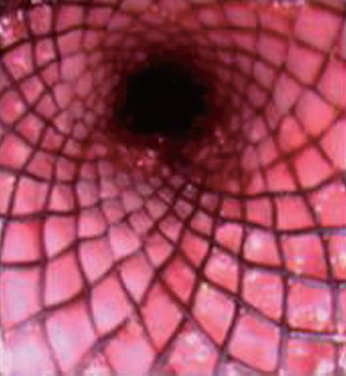
Figure 1 Proximal view of a fully opened stent.
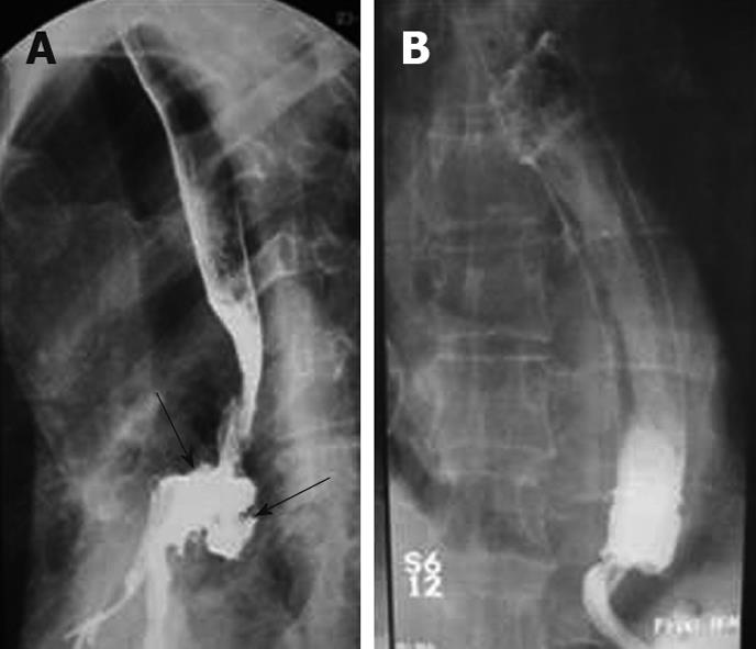
Figure 2 Palliation of tracheoesophageal fistula with stenting.
A: Barium esophagogram showing a tracheo-esophageal fistula resulting from lung cancer; B: Complete occlusion of the fistula after stenting. Arrows show a large tracheoesophageal fistula.
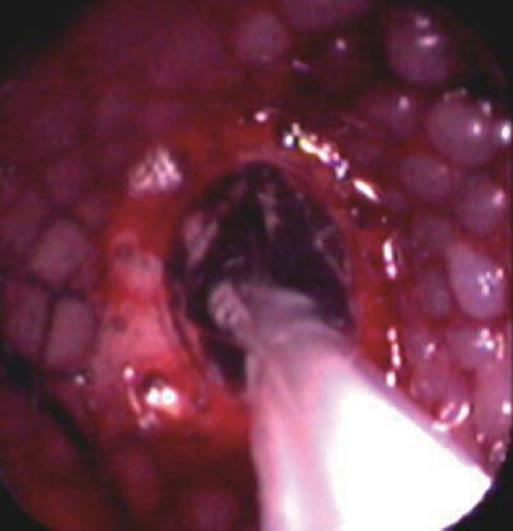
Figure 3 In some cases with insufficient stent expansion due to tight stricture, endoscopic balloon dilatation was performed through the opened stent.
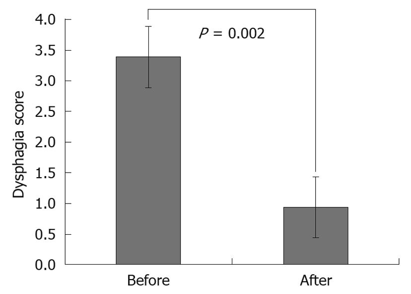
Figure 4 Comparison of oral alimentation status before and after placement of self expandable metallic stents.
Figure shows the change in dysphagia score on day 3 after stenting. For the scoring system, see Materials and Methods section.
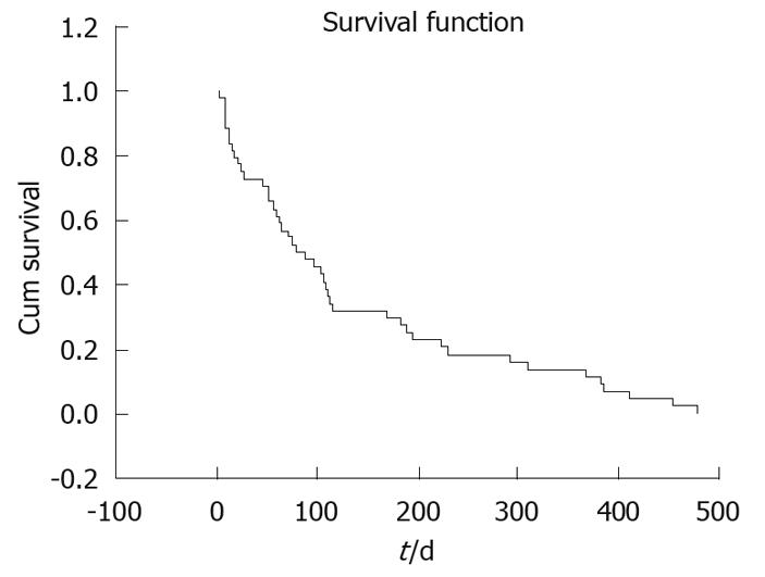
Figure 5 Kaplan-Meier survival curve of 90 patients following stenting.
- Citation: Dobrucali A, Caglar E. Palliation of malignant esophageal obstruction and fistulas with self expandable metallic stents. World J Gastroenterol 2010; 16(45): 5739-5745
- URL: https://www.wjgnet.com/1007-9327/full/v16/i45/5739.htm
- DOI: https://dx.doi.org/10.3748/wjg.v16.i45.5739


