Copyright
©2010 Baishideng Publishing Group Co.
World J Gastroenterol. Nov 14, 2010; 16(42): 5391-5394
Published online Nov 14, 2010. doi: 10.3748/wjg.v16.i42.5391
Published online Nov 14, 2010. doi: 10.3748/wjg.v16.i42.5391
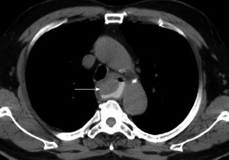
Figure 1 Computed tomography revealing a slight hyperdense tumor lesion (arrow) with a smooth surface over the upper esophagus, and no false lumen.
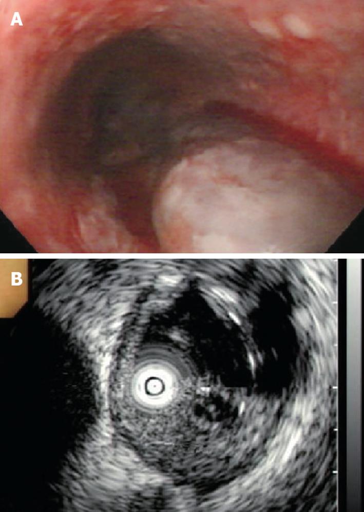
Figure 2 Esophagogastroduodenoscopy revealing a purplish bleeding esophageal submucosal tumor (A) and endoscopic ultrasound revealing a mixed echoic tumor with an anechoic component and an irregular at the submucosal layer (B).
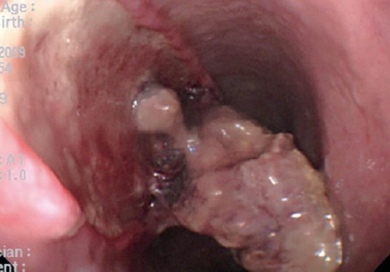
Figure 3 Follow-up esophagogastroduodenoscopy 2 d later revealing a dissected necrotic mass lesion with ulcer formation.
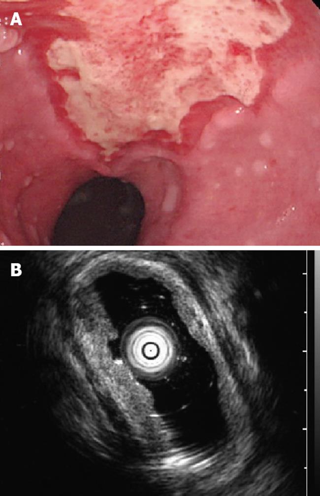
Figure 4 Esophagogastroduodenoscopy revealing a shallow ulcer without a tumor (A) and endoscopic ultrasound revealing thickening mucosal and submucosal layers, an intact muscularis propria layer, and no submucosal tumor (B) during the follow-up one week later.
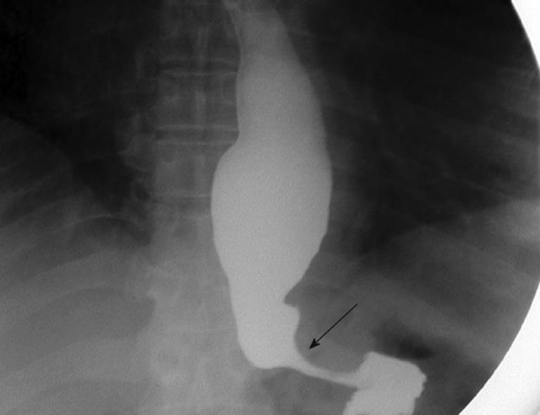
Figure 5 Esophagography revealing a dilated esophageal lumen with smooth narrowing of the esophagocardiac junction (arrow).
- Citation: Chu YY, Sung KF, Ng SC, Cheng HT, Chiu CT. Achalasia combined with esophageal intramural hematoma: Case report and literature review. World J Gastroenterol 2010; 16(42): 5391-5394
- URL: https://www.wjgnet.com/1007-9327/full/v16/i42/5391.htm
- DOI: https://dx.doi.org/10.3748/wjg.v16.i42.5391


