Copyright
©2010 Baishideng Publishing Group Co.
World J Gastroenterol. Nov 14, 2010; 16(42): 5280-5285
Published online Nov 14, 2010. doi: 10.3748/wjg.v16.i42.5280
Published online Nov 14, 2010. doi: 10.3748/wjg.v16.i42.5280
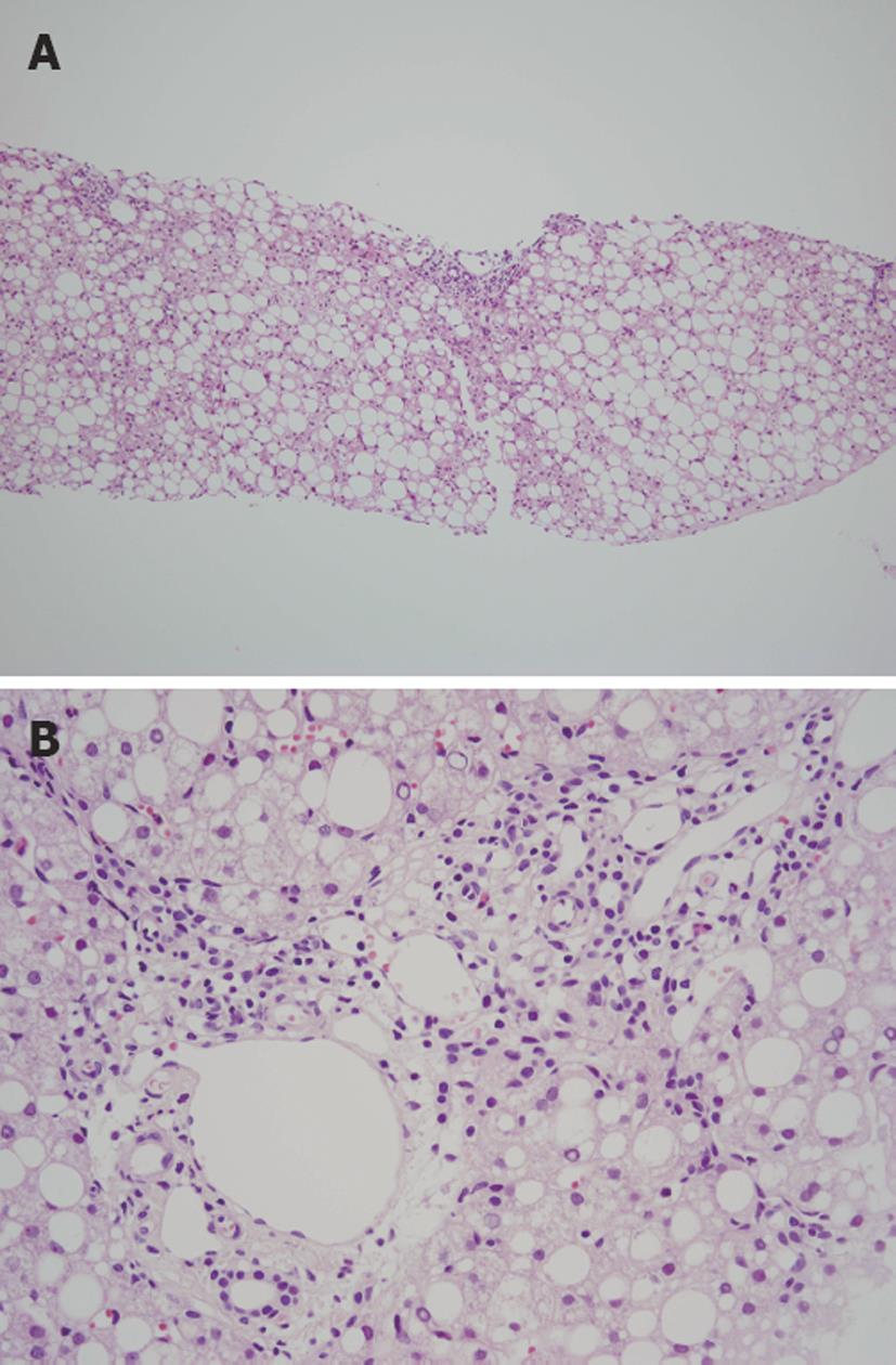
Figure 1 Histological appearance of a case of pediatric nonalcoholic fatty liver disease.
A: Marked steatosis with panlobular distribution is observed (HE stain, × 40); B: Moderate inflammation is observed in the portal area (HE stain, × 200).
- Citation: Takahashi Y, Fukusato T. Pediatric nonalcoholic fatty liver disease: Overview with emphasis on histology. World J Gastroenterol 2010; 16(42): 5280-5285
- URL: https://www.wjgnet.com/1007-9327/full/v16/i42/5280.htm
- DOI: https://dx.doi.org/10.3748/wjg.v16.i42.5280


