Copyright
©2010 Baishideng Publishing Group Co.
World J Gastroenterol. Oct 28, 2010; 16(40): 5111-5117
Published online Oct 28, 2010. doi: 10.3748/wjg.v16.i40.5111
Published online Oct 28, 2010. doi: 10.3748/wjg.v16.i40.5111
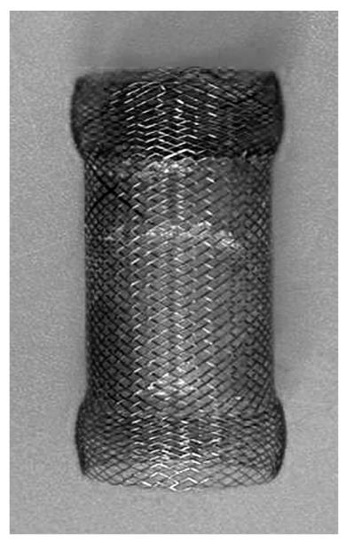
Figure 1 Photograph of a partially covered self-expanding metallic stent.
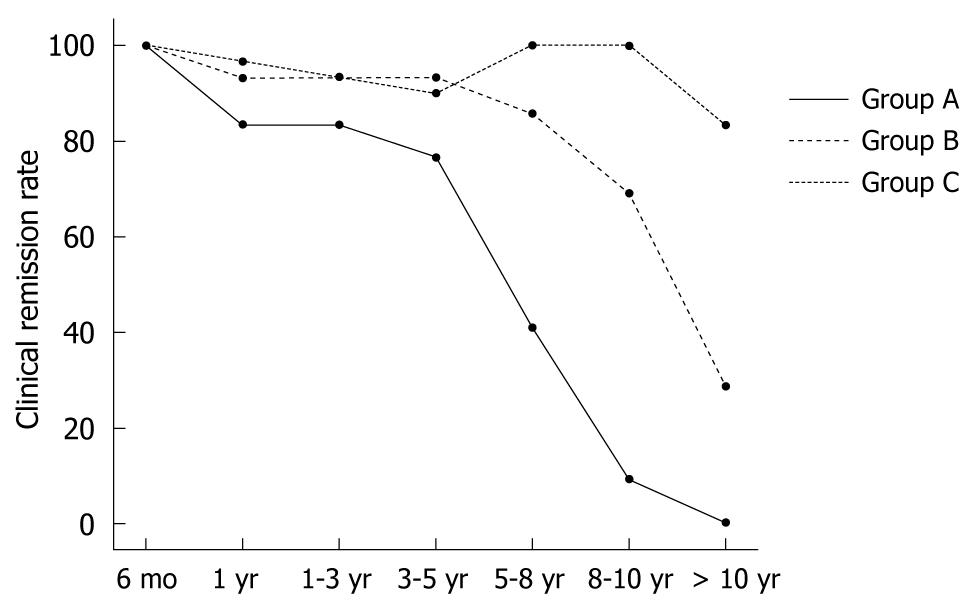
Figure 2 Clinical remission rates in comparison with self-expanding metallic stents with a diameter of 30 mm (Group C), 25 mm (Group B) or 20 mm (Group A).
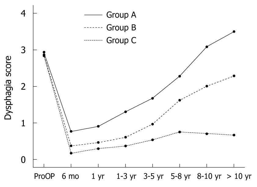
Figure 3 Dysphagia scores among the three groups before self-expanding metallic stents placement at different follow-up time intervals.
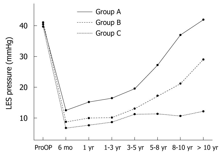
Figure 4 Lower esophageal sphincter pressures assessed by manometry among the three groups before self-expanding metallic stent placement at different follow-up time intervals.
LES: Lower esophageal sphincter.
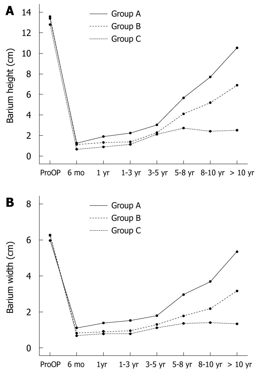
Figure 5 The barium height (A) and width (B) assessed by a timed barium esophagram among the three groups before self-expanding metallic stent placement at different follow-up time intervals.
- Citation: Cheng YS, Ma F, Li YD, Chen NW, Chen WX, Zhao JG, Wu CG. Temporary self-expanding metallic stents for achalasia: A prospective study with a long-term follow-up. World J Gastroenterol 2010; 16(40): 5111-5117
- URL: https://www.wjgnet.com/1007-9327/full/v16/i40/5111.htm
- DOI: https://dx.doi.org/10.3748/wjg.v16.i40.5111


