Copyright
©2010 Baishideng Publishing Group Co.
World J Gastroenterol. Oct 7, 2010; 16(37): 4704-4708
Published online Oct 7, 2010. doi: 10.3748/wjg.v16.i37.4704
Published online Oct 7, 2010. doi: 10.3748/wjg.v16.i37.4704
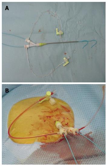
Figure 1 Y-shaped sheath and catheter systems.
A: Y-shaped sheath with 2 valves and 2 catheters (3.2 French in size) through the angiographic sheath. B: Placing Y-shaped sheath introducers through the right femoral artery.
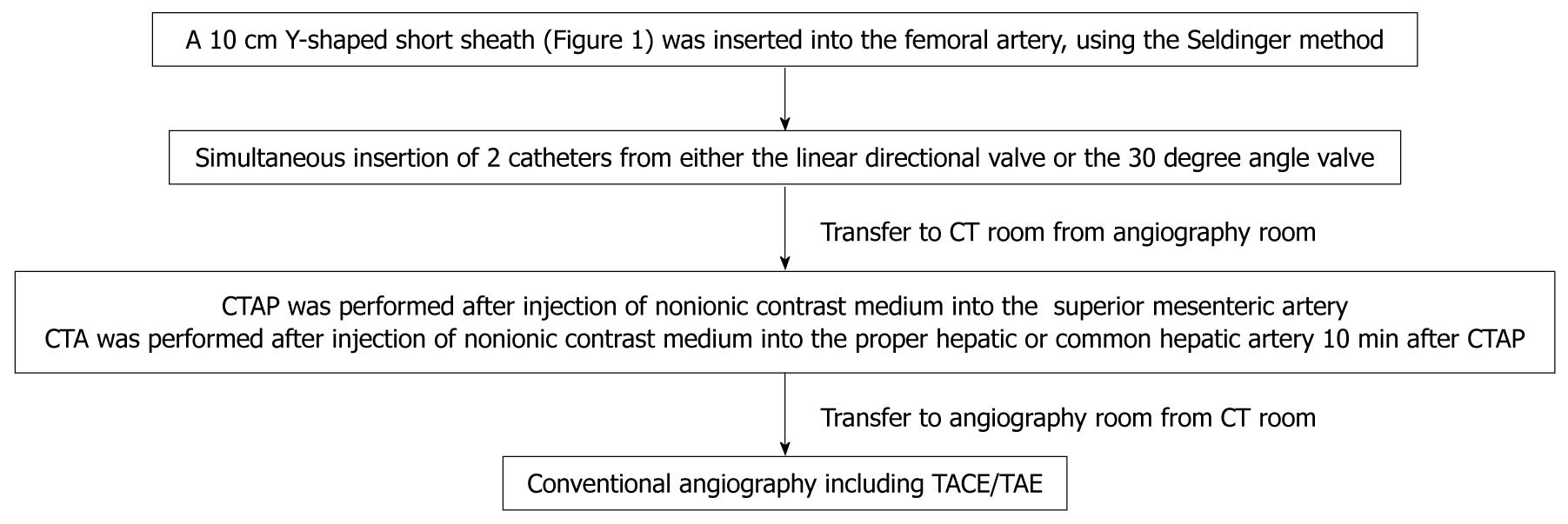
Figure 2 The whole diagnostic process including insertion of the catheters, angiography, computed tomography arterial portography and computed tomography arteriography.
CT: Computed tomography; CTAP: CT arterial portography; CTA: CT arteriography; TACE: Transcatheter arterial chemoembolization; TAE: Transarterial embolization.
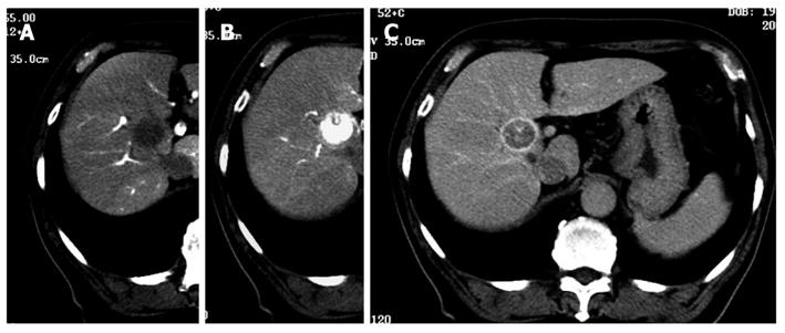
Figure 3 Representative case of classical hepatocellular carcinoma diagnosed using 3.
2 Fr catheters in the Y-shaped sheath. A: Computed tomography (CT) arterial portography; B: CT arteriography early phase; C: CT arteriography late phase.
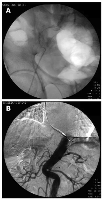
Figure 4 Sheath insertion was difficult and insertion of the 3.
2 Fr catheter was impossible. A: Representative case with strong curvature of the femoral artery; B: Representative case with strong curvature of the abdominal aorta.
- Citation: Ishikawa T, Higuchi K, Kubota T, Seki KI, Honma T, Yoshida T, Nemoto T, Takeda K, Kamimura T. Usefulness of Y-shaped sheaths in CT angiography for examination of liver tumors. World J Gastroenterol 2010; 16(37): 4704-4708
- URL: https://www.wjgnet.com/1007-9327/full/v16/i37/4704.htm
- DOI: https://dx.doi.org/10.3748/wjg.v16.i37.4704


