Copyright
©2010 Baishideng Publishing Group Co.
World J Gastroenterol. Oct 7, 2010; 16(37): 4661-4669
Published online Oct 7, 2010. doi: 10.3748/wjg.v16.i37.4661
Published online Oct 7, 2010. doi: 10.3748/wjg.v16.i37.4661
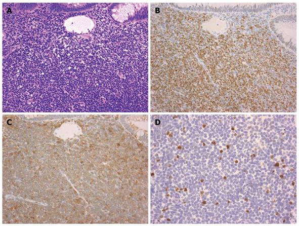
Figure 1 Histological features of mantle cell lymphoma.
A: Rectal biopsied specimen demonstrated monomorphic proliferation of medium-sized lymphoid cells (Hematoxylin and eosin staining). This case illustrated typical immunohistochemical features of mantle cell lymphoma, showing positivity for cyclin D1 and CD5 staining; B: Immunohistochemical staining for cyclin D1; C: Immunohistochemical staining for CD5; D: Approximately 5% of neoplastic cells were positive for Ki-67 staining; consequently, this case was classified as a low mitotic rate. A-C: Original magnification, × 200; D: × 400.
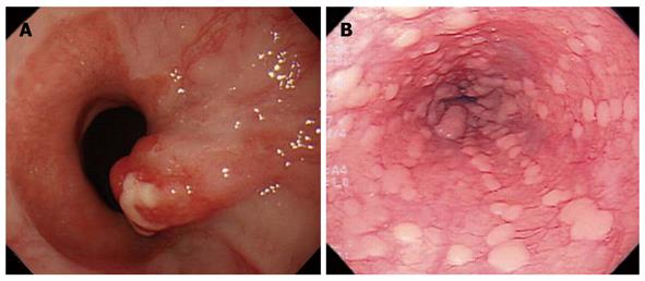
Figure 2 Esophageal lesions of mantle cell lymphoma.
A: A protruded tumor was seen in the esophagogastric junction; B: In another patient, multiple whitish plaques were seen throughout the whole esophagus.
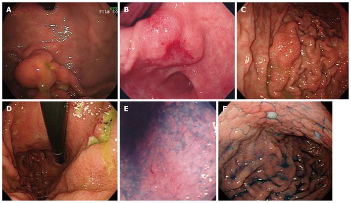
Figure 3 Gastric lesions of mantle cell lymphoma.
A: Protruded type (n = 6) often resembled submucosal tumor; B: One patient with a protruded type lesion was diagnosed with stage I primary gastrointestinal mantle cell lymphoma; C: Fold thickening type (n = 6); D: Ulcerative type (n = 6); E: In one patient, only a change in mucosal color with redness was seen. This case was classified as the superficial type (n = 7); F: Among superficial type lesions, cobblestone-like mucosa with a few shallow ulcers was also seen.
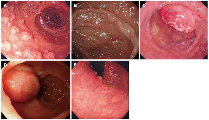
Figure 4 Intestinal lesions of mantle cell lymphoma.
From the duodenum to the rectum, multiple lymphomatous polyposis (MLP) was dominant (n = 17). A, B: Typical features of MLP were diffuse multiple micropolyps; C: Some large tumorous polyps were sometimes found together with micropolyps; D: Protruded type (n = 4); E: Superficial type (n = 1, arrow).
Figure 5 Overall survival (A) and event-free survival (B) according to administration of the hyper-CVAD/MA regimen.
1Log-rank test.
- Citation: Iwamuro M, Okada H, Kawahara Y, Shinagawa K, Morito T, Yoshino T, Yamamoto K. Endoscopic features and prognoses of mantle cell lymphoma with gastrointestinal involvement. World J Gastroenterol 2010; 16(37): 4661-4669
- URL: https://www.wjgnet.com/1007-9327/full/v16/i37/4661.htm
- DOI: https://dx.doi.org/10.3748/wjg.v16.i37.4661


