Copyright
copy;2010 Baishideng Publishing Group Co.
World J Gastroenterol. Sep 28, 2010; 16(36): 4515-4518
Published online Sep 28, 2010. doi: 10.3748/wjg.v16.i36.4515
Published online Sep 28, 2010. doi: 10.3748/wjg.v16.i36.4515
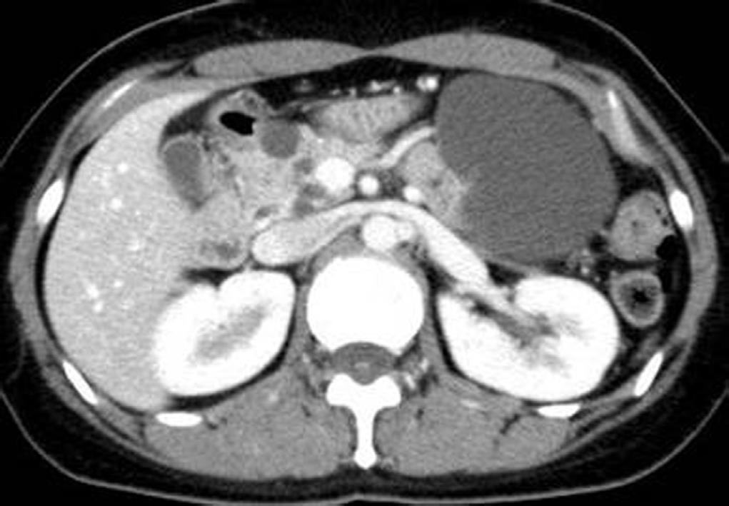
Figure 1 Abdominal computed tomography shows several cystic lesions in the pancreas (32-year-old female).
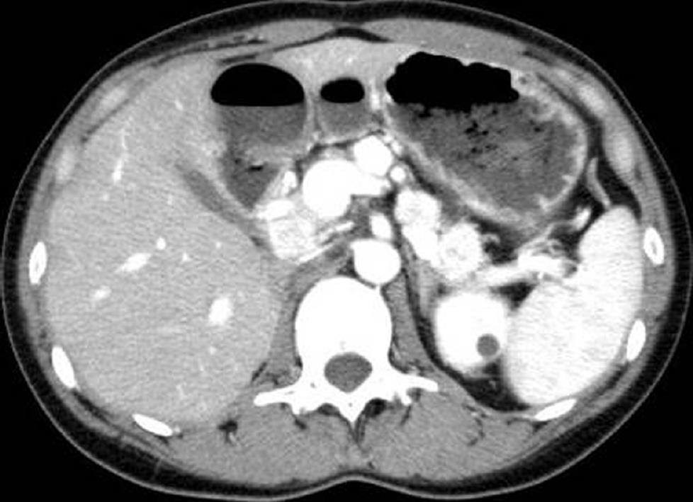
Figure 2 Contrast-enhanced abdominal computed tomography reveals several pancreatic mass lesions that are strongly enhanced (33-year-old female)[14].
- Citation: Tamura K, Nishimori I, Ito T, Yamasaki I, Igarashi H, Shuin T. Diagnosis and management of pancreatic neuroendocrine tumor in von Hippel-Lindau disease. World J Gastroenterol 2010; 16(36): 4515-4518
- URL: https://www.wjgnet.com/1007-9327/full/v16/i36/4515.htm
- DOI: https://dx.doi.org/10.3748/wjg.v16.i36.4515


