Copyright
©2010 Baishideng Publishing Group Co.
World J Gastroenterol. Sep 21, 2010; 16(35): 4491-4493
Published online Sep 21, 2010. doi: 10.3748/wjg.v16.i35.4491
Published online Sep 21, 2010. doi: 10.3748/wjg.v16.i35.4491
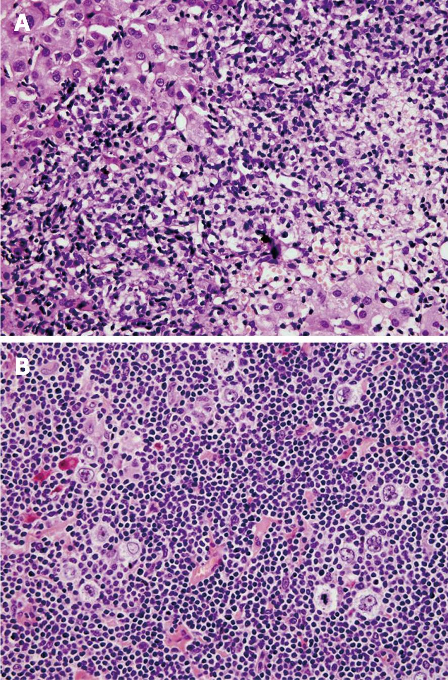
Figure 1 Nodular lymphocyte-predominant Hodgkin lymphoma of the liver and axillary lymph node.
A: A portal tract showing a dense lymphoid infiltrate. The multiple sections revealed just a few scattered, CD20 positive neoplastic cells (HE stain, × 40); B: The popcorn (Hodgkin lymphoma) cells with the typically lobulated nuclei in a background of small lymphoid cells (HE stain, × 40).
- Citation: Mrzljak A, Gasparov S, Kardum-Skelin I, Colic-Cvrlje V, Ostojic-Kolonic S. Febrile cholestatic disease as an initial presentation of nodular lymphocyte-predominant Hodgkin lymphoma. World J Gastroenterol 2010; 16(35): 4491-4493
- URL: https://www.wjgnet.com/1007-9327/full/v16/i35/4491.htm
- DOI: https://dx.doi.org/10.3748/wjg.v16.i35.4491


