Copyright
©2010 Baishideng Publishing Group Co.
World J Gastroenterol. Sep 14, 2010; 16(34): 4363-4366
Published online Sep 14, 2010. doi: 10.3748/wjg.v16.i34.4363
Published online Sep 14, 2010. doi: 10.3748/wjg.v16.i34.4363
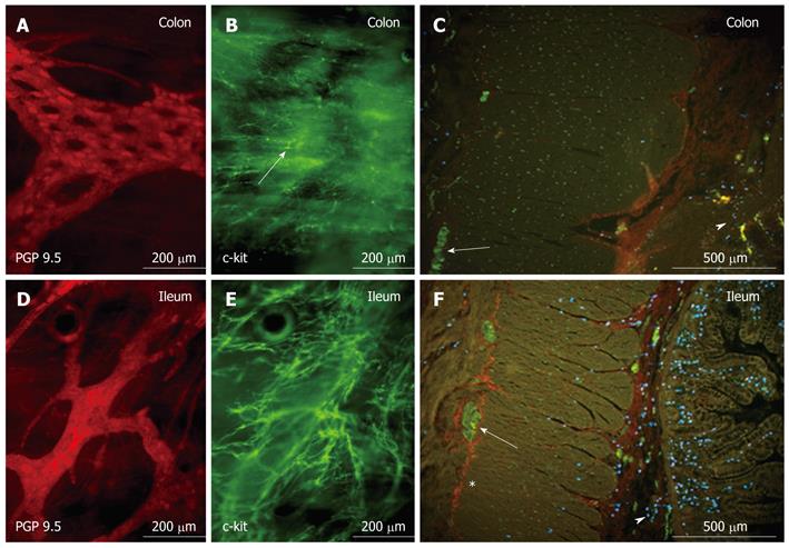
Figure 1 Whole mount preparation of the myenteric plexus of the patient’s colon (A and B) and ileum (D and E), which shows abnormal morphology (arrow in B) and extreme low density of c-kit-positive interstitial cells of Cajal in the colonic biopsy specimen (B), while a dense interstitial cells of Cajal network with normal cell morphology was present in the ileum (E).
Also remarkable are the holes in the colonic ganglia (A), which are very uncommon in young patients. D shows normal morphology and density of ganglia and protein gene product (PGP)-positive myenteric neurons in the ileum. Cross-sections of colon (C) and ileum (F) illustrate the normal appearance of myenteric interstitial cells of Cajal (ICCs) (*c-kit, red) in the ileum, but the complete absence of myenteric ICCs in the colon. (A and D: staining for the neuronal marker PGP 9.5, red; PH164, The Binding Site, Birmingham, UK; B and E: staining for c-kit as ICC marker, green; PC34, Oncogene, Boston, MA, USA; C and F: PGP-positive neurons (green) in a ganglion (arrow), mast cells in mucosa/submucosa (arrowhead), stained by a tryptase antibody, blue; MAB1222, Chemicon, Schwalbach, Germany.
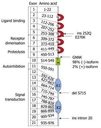
Figure 2 Schematic representation of the molecular and functional structure of the tyrosine kinase Kit (CD117), which indicated the patient’s mutations in the deduced amino acid sequence.
Details are discussed in the text (Ig, immunoglobulin-like domain; TM, transmembranous domain; K1, ATP-binding region of kinase domain; KI, kinase-insert-region; K2, region of phosphorylation of kinase domain).
- Citation: Breuer C, Oh J, Molderings GJ, Schemann M, Kuch B, Mayatepek E, Adam R. Therapy-refractory gastrointestinal motility disorder in a child with c-kit mutations. World J Gastroenterol 2010; 16(34): 4363-4366
- URL: https://www.wjgnet.com/1007-9327/full/v16/i34/4363.htm
- DOI: https://dx.doi.org/10.3748/wjg.v16.i34.4363


