Copyright
copy;2010 Baishideng Publishing Group Co.
World J Gastroenterol. Sep 7, 2010; 16(33): 4233-4236
Published online Sep 7, 2010. doi: 10.3748/wjg.v16.i33.4233
Published online Sep 7, 2010. doi: 10.3748/wjg.v16.i33.4233
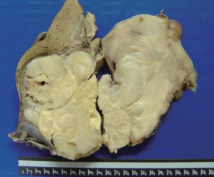
Figure 1 Well circumscribed and nodular mass, tan-white.
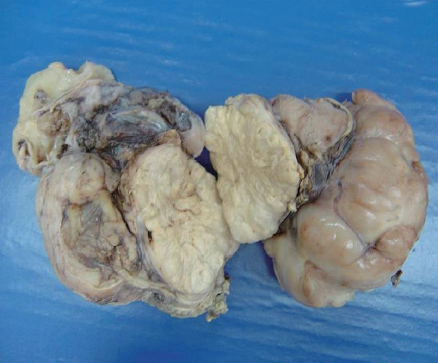
Figure 2 Multinodular mass, tan-white and shining.
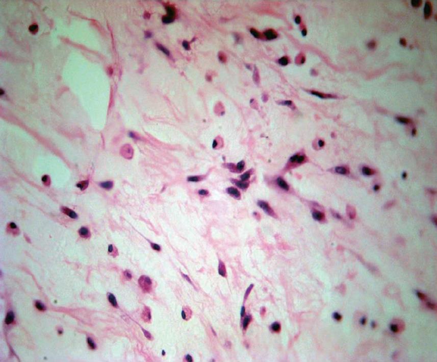
Figure 3 Spindle and inflammatory cells in a myxoid background (HE stain, × 100).
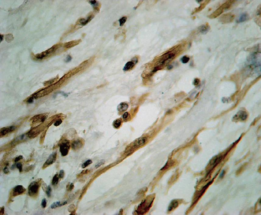
Figure 4 Smooth muscle actin positive in the spindle cell tumor (immunostaining, × 200).
- Citation: Pannain VL, Passos JV, Filho ADR, Villela-Nogueira C, Caroli-Bottino A. Agressive inflammatory myofibroblastic tumor of the liver with underlying schistosomiasis: A case report. World J Gastroenterol 2010; 16(33): 4233-4236
- URL: https://www.wjgnet.com/1007-9327/full/v16/i33/4233.htm
- DOI: https://dx.doi.org/10.3748/wjg.v16.i33.4233


