Copyright
copy;2010 Baishideng Publishing Group Co.
World J Gastroenterol. Sep 7, 2010; 16(33): 4180-4186
Published online Sep 7, 2010. doi: 10.3748/wjg.v16.i33.4180
Published online Sep 7, 2010. doi: 10.3748/wjg.v16.i33.4180
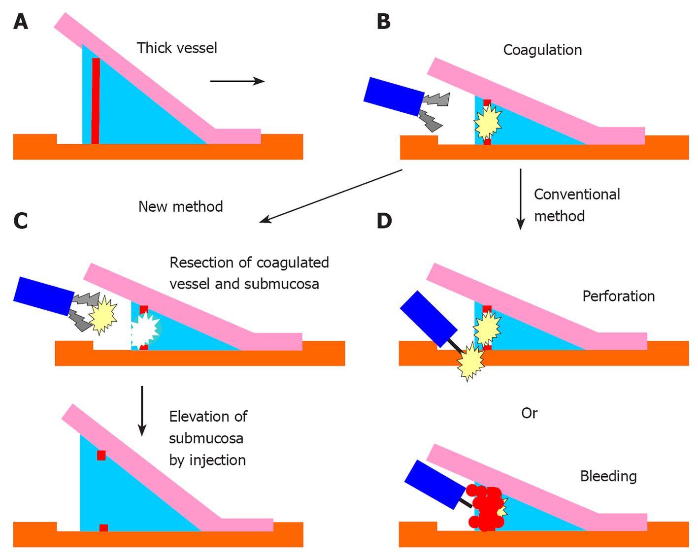
Figure 1 Schema of the efficacy of the new hemostatic forceps method.
A: A thick vessel is detected during the submucosal dissection; B: The thick vessel is coagulated using the hemostatic forceps. The vessel and surrounding submucosa become whitish, and the submucosa becomes less elevated; C: In the new method, the coagulated vessel is removed using the hemostatic forceps and the surrounding submucosa is also dissected. After the treatment, the submucosa is elevated by injecting it with hyaluronic acid and glycerin; D: In the conventional use of hemostatic forceps, perforation can occur because of inadequate submucosal elevation; moreover, hemorrhage can occur from the coagulated vessel while dissecting it with a knife.
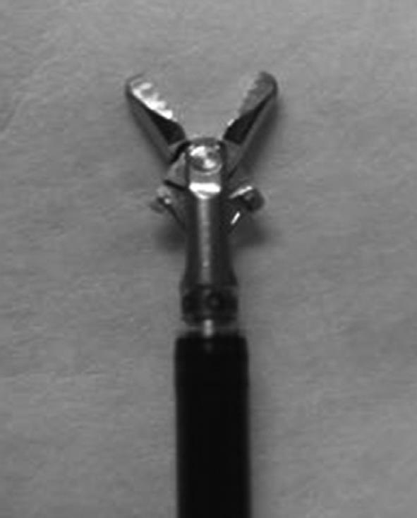
Figure 2 The hemostatic forceps, coagrasper.
These forceps have a rotation function and small teeth.
Figure 3 Clinical use of the new method in endoscopic submucosal dissection for colorectal tumors.
A: A vessel with a diameter of 1 mm was detected; B: It was coagulated in the soft coagulation mode. Thereafter, the vessel and the surrounding submucosa became whitish, and the submucosa became less elevated. Submucosal dissection with a knife was slightly difficult because of the obscured view of the submucosa and the proximity of the muscularis propria; C: The whitish coagulated vessel was removed using the hemostatic forceps in the “endocut” mode. Subsequently, the whitish surrounding submucosa was dissected using the forceps. Thereafter, the view of the submucosa was improved.
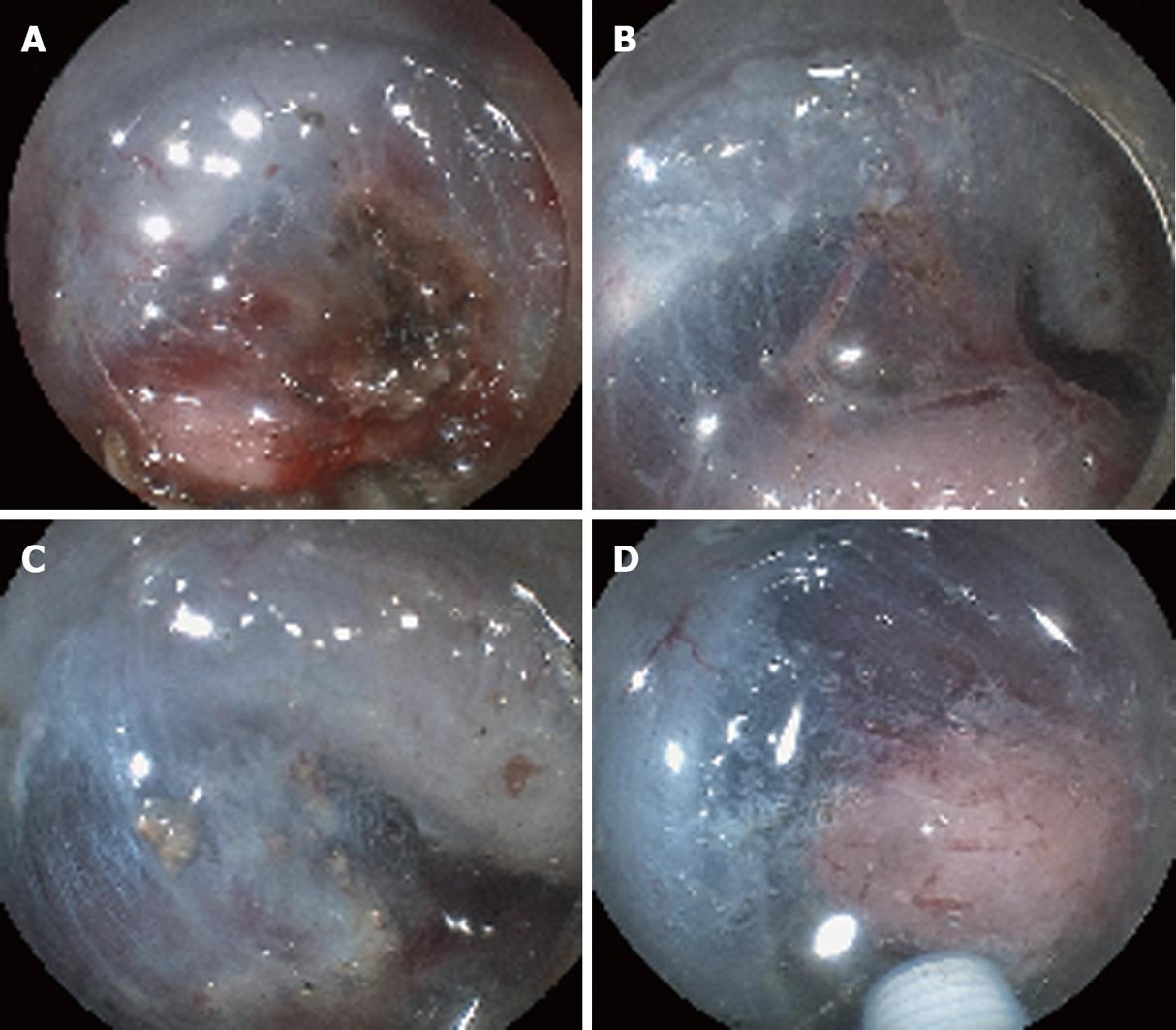
Figure 4 Clinical use of the new method for hemorrhage from the submucosal vessel.
A: Coagulation of the hemorrhagic vessels made the vessels and the surrounding submucosa brownish; B: The brownish vessel was removed and the surrounding submucosa was dissected. The view of the submucosa became clear and the other vessel could be detected; C: The vessel was coagulated using the forceps to prevent perioperative hemorrhage; D: The coagulated vessel was removed and the surrounding submucosa was dissected. Thereafter, the view of the submucosa was improved.
Figure 5 Clinical use of the new method for brownish coagulated submucosa.
A: Brownish coagulated submucosa was detected; however, the view of the submucosa was obscured; B: The brownish submucosa was dissected using the new method; C: The view of the submucosa was restored.
- Citation: Yoshida N, Naito Y, Kugai M, Inoue K, Wakabayashi N, Yagi N, Yanagisawa A, Yoshikawa T. Efficient hemostatic method for endoscopic submucosal dissection of colorectal tumors. World J Gastroenterol 2010; 16(33): 4180-4186
- URL: https://www.wjgnet.com/1007-9327/full/v16/i33/4180.htm
- DOI: https://dx.doi.org/10.3748/wjg.v16.i33.4180


