Copyright
copy;2010 Baishideng Publishing Group Co.
World J Gastroenterol. Sep 7, 2010; 16(33): 4164-4168
Published online Sep 7, 2010. doi: 10.3748/wjg.v16.i33.4164
Published online Sep 7, 2010. doi: 10.3748/wjg.v16.i33.4164
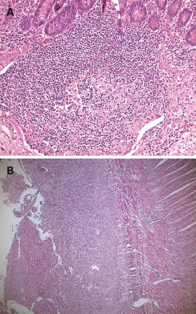
Figure 1 Histopathologic view of typhoid lesions.
A: Typhoid nodule, there are macrophages containing bacteria, red blood cells, and nuclear debris from small nodular aggregates in Peyer’s patches (HE stain, × 20 objective); B: Typhoid ulceration (HE stain, × 5 objective).
- Citation: Sümer A, Kemik &, Dülger AC, Olmez A, Hasirci I, Kişli E, Bayrak V, Bulut G, Kotan &. Outcome of surgical treatment of intestinal perforation in typhoid fever. World J Gastroenterol 2010; 16(33): 4164-4168
- URL: https://www.wjgnet.com/1007-9327/full/v16/i33/4164.htm
- DOI: https://dx.doi.org/10.3748/wjg.v16.i33.4164


