Copyright
©2010 Baishideng.
World J Gastroenterol. Jan 21, 2010; 16(3): 289-295
Published online Jan 21, 2010. doi: 10.3748/wjg.v16.i3.289
Published online Jan 21, 2010. doi: 10.3748/wjg.v16.i3.289
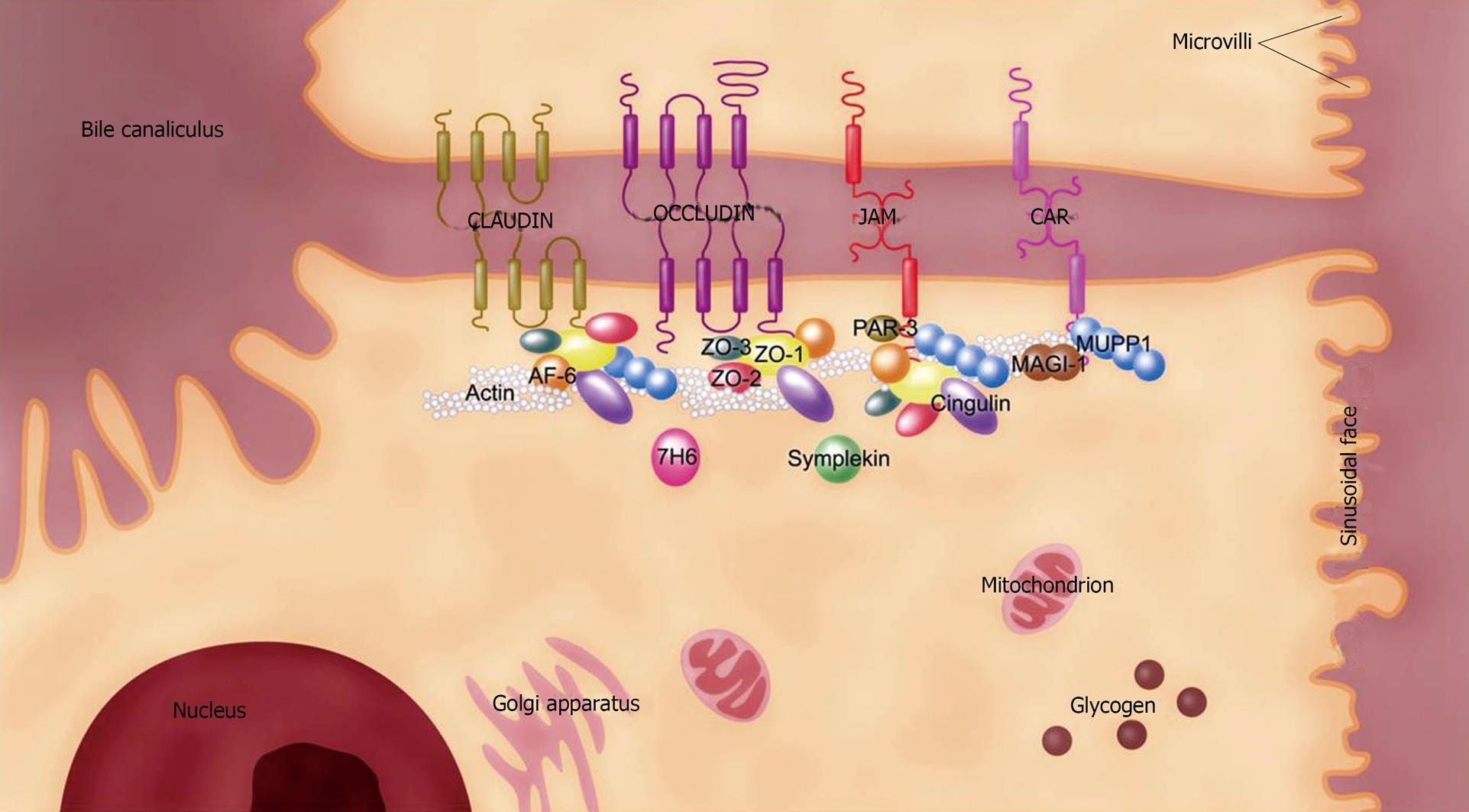
Figure 1 Molecular structure of the TJ in the mammalian liver.
The TJ in the liver is associated with hepatocytes and bile duct cells. TJ location around the bile canaliculus between 2 adjacent hepatocytes is shown. For simplicity, the molecular structure depicted in this figure represents the TJ molecules found in the mammalian liver. Claudin, occludin, JAM, and CAR are 4 core units for constituting TJ by uniting a panel of peripheral proteins like ZO-1 to form multiprotein complexes. TJ molecules display differential localizations in the mammalian liver, such that some of them like human symplekin and mouse CAR-2 are associated with both hepatocytes and bile duct cells while others, such as mouse CAR-1, are only found in the latter cell type. CAR: Coxsackievirus and adenovirus receptor; JAM: Junctional adhesion molecule; MAGI-1: Membrane-associated guanylate kinase inverted-1; MUPP1: Multiple PDZ domain protein-1; PAR-3: Partitioning defective 3 homolog; TJ: Tight junction; ZO: Zonula occludens.
Figure 2 The TJ at different stages of liver cancer progression and distant liver metastasis.
During viral infection, HCV binds onto several TJ molecules (claudin-1 and occludin) and co-receptors (CD81 and SR-BI) on hepatocytes before its internalization. This event leads to hepatic steatosis and/or cirrhosis before the subsequent development of primary liver cancer that is associated with TJ deregulation. For metastatic liver cancer originating from the intrahepatic site or distant organs such as breast and colorectum, there is an initial loss of TJ molecules at the primary tumor site and a subsequent gain of these molecules in the liver with tumor cell colonization. HCV: Hepatitis C virus; SR-BI: Scavenger receptor class B member-I.
- Citation: Lee NP, Luk JM. Hepatic tight junctions: From viral entry to cancer metastasis. World J Gastroenterol 2010; 16(3): 289-295
- URL: https://www.wjgnet.com/1007-9327/full/v16/i3/289.htm
- DOI: https://dx.doi.org/10.3748/wjg.v16.i3.289


