Copyright
©2010 Baishideng.
World J Gastroenterol. Aug 7, 2010; 16(29): 3680-3686
Published online Aug 7, 2010. doi: 10.3748/wjg.v16.i29.3680
Published online Aug 7, 2010. doi: 10.3748/wjg.v16.i29.3680
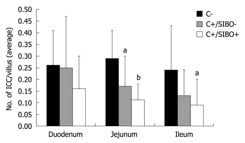
Figure 1 The number of interstitial cells of Cajal per villus in the duodenum, jejunum, and ileum.
C-: Rats that received vehicle gavage; C+/SIBO-: Rats gavaged with Campylobacter but which did not develop small intestinal bacterial overgrowth (SIBO); C+/SIBO+: Rats gavaged with Campylobacter that later developed SIBO. aP < 0.05, bP < 0.001 vs control.
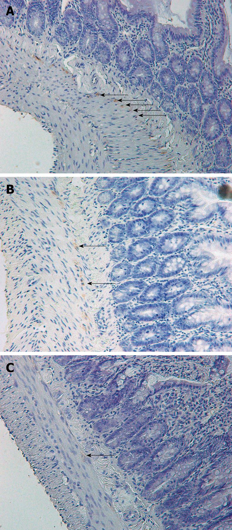
Figure 2 Interstitial cells of Cajal (arrows) in the ileum stained with CD117 (magnification 40 ×).
A: Control rat; B: Rat exposed to Campylobacter that did not develop small intestinal bacterial overgrowth (SIBO), demonstrating persistent CD117 staining of interstitial cells of Cajal (ICC) cells; C: Rat exposed to Campylobacter that developed SIBO, demonstrating a reduction in CD117 staining of ICC cells. In this case, the staining is slight and the arrow indicates a “possible” stained cell.
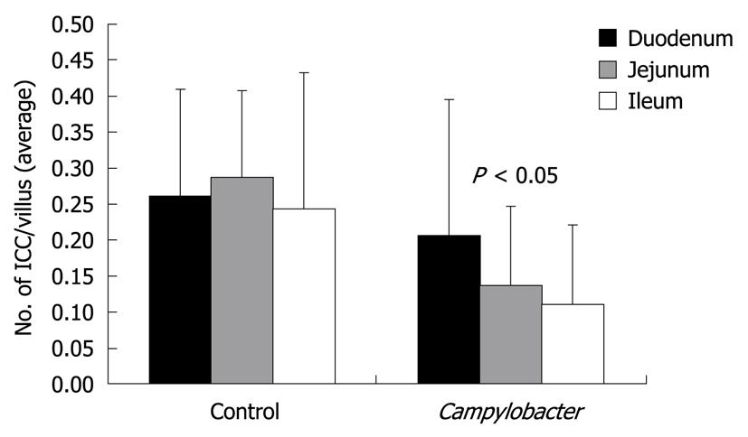
Figure 3 The effect of Campylobacter on interstitial cells of Cajal is greater in the distal small bowel (based on one-way analysis of variance).
- Citation: Jee SR, Morales W, Low K, Chang C, Zhu A, Pokkunuri V, Chatterjee S, Soffer E, Conklin JL, Pimentel M. ICC density predicts bacterial overgrowth in a rat model of post-infectious IBS. World J Gastroenterol 2010; 16(29): 3680-3686
- URL: https://www.wjgnet.com/1007-9327/full/v16/i29/3680.htm
- DOI: https://dx.doi.org/10.3748/wjg.v16.i29.3680


