Copyright
©2010 Baishideng.
World J Gastroenterol. Jul 14, 2010; 16(26): 3339-3342
Published online Jul 14, 2010. doi: 10.3748/wjg.v16.i26.3339
Published online Jul 14, 2010. doi: 10.3748/wjg.v16.i26.3339
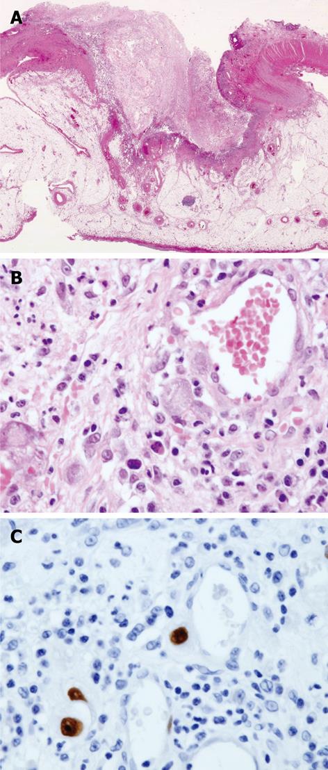
Figure 1 Histological examination of perforated ileum.
A: Perforated ileal lesion (hematoxylin and eosin stain) × 40; B: × 400; C: Cytomegalovirus infection was evident in the same lesion, by immunohistochemical staining with anti-cytomegalovirus antibody (× 200).
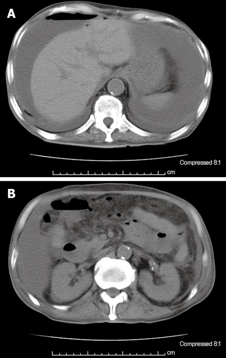
Figure 2 Abdominal computed tomography.
A: Free-air, ascites; B: Swelling of the ileal loop.
- Citation: Sano S, Ueno H, Yamagami K, Yakushiji Y, Isaka Y, Kawasaki I, Takemura M, Inoue T, Hosoi M. Isolated ileal perforation due to cytomegalovirus reactivation during management of terbinafine hypersensitivity. World J Gastroenterol 2010; 16(26): 3339-3342
- URL: https://www.wjgnet.com/1007-9327/full/v16/i26/3339.htm
- DOI: https://dx.doi.org/10.3748/wjg.v16.i26.3339


