Copyright
©2010 Baishideng.
World J Gastroenterol. Jun 28, 2010; 16(24): 3078-3082
Published online Jun 28, 2010. doi: 10.3748/wjg.v16.i24.3078
Published online Jun 28, 2010. doi: 10.3748/wjg.v16.i24.3078
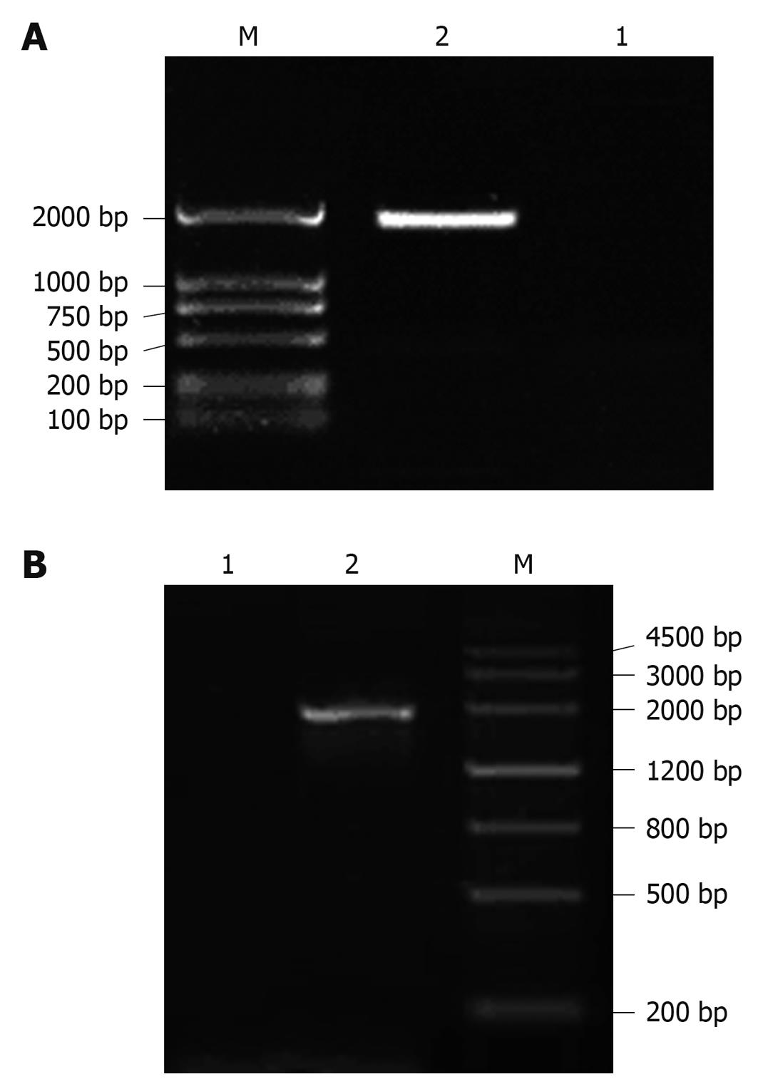
Figure 1 Characterization of polymerase chain reaction (PCR) amplification by electrophoresis.
A: Amplification of calreticulin (CRT)/hepatitis B surface antigen (HBsAg) fusion gene by PCR. Lane 1: Negative control; Lane 2: CRT/HBsAg fusion gene recombinant pJW4303 plasmid; Lane M: DNA marker; B: Characterization of CRT/HBsAg fusion gene recombinant pENTR/D-TOPO transfer vector by PCR. Lane 1: Negative control; Lane 2: CRT/HBsAg fusion gene recombinant pENTR/D-TOPO transfer vector amplified by specific PCR; Lane M: DNA marker.
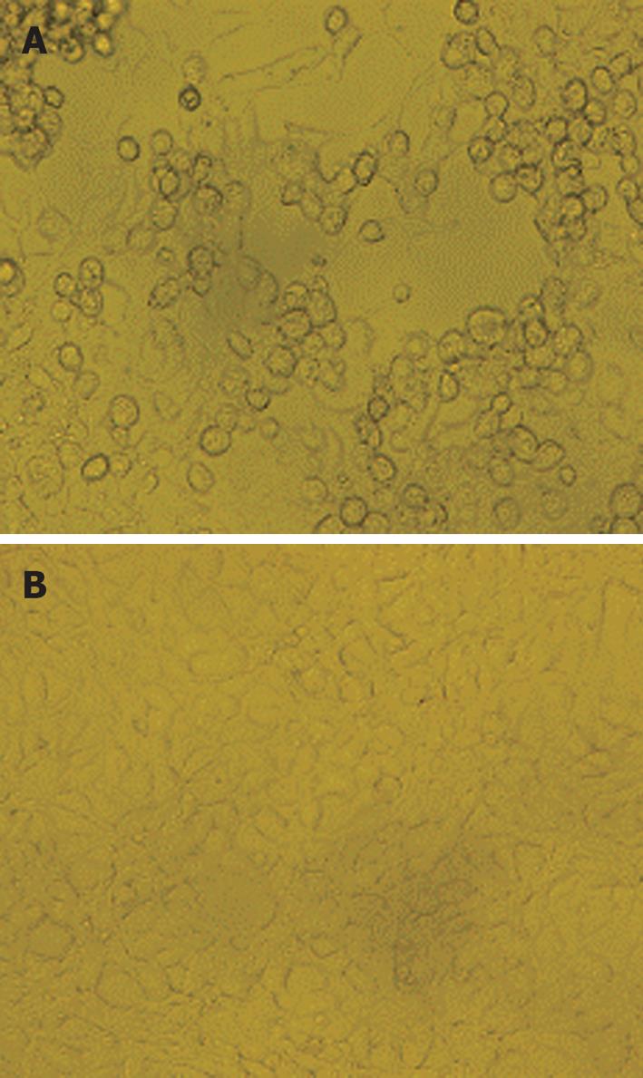
Figure 2 Culture of recombinant adenovirus in HEK293A cells.
A: CPE was observed in recombinant adenovirus-containing HEK293A cells; B: Normal HEK293A cells cultured as control.
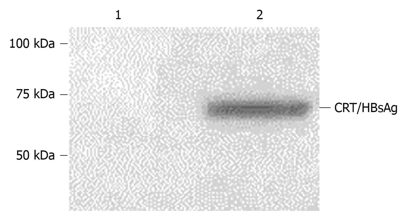
Figure 3 Characterization of CRT/HBsAg fusion protein expression by Western blotting.
The expression of CRT/HBsAg fusion protein in 293A cells was determined in 293A cells transfected with Ad-CRT/HBsAg and Ad-LacZ by Western blotting analysis. Human anti-HBsAg positive serum was used at a 1:100 dilution for the detection of CRT/HBsAg fusion protein expression. Lane 1: Lysate from 293 cells transfected with Ad-LacZ; Lane 2: Lysate from 293 cells transfected with Ad-CRT/HBsAg.
- Citation: Ma CL, Wang GB, Gu RG, Wang F. Construction and characterization of calreticulin-HBsAg fusion gene recombinant adenovirus expression vector. World J Gastroenterol 2010; 16(24): 3078-3082
- URL: https://www.wjgnet.com/1007-9327/full/v16/i24/3078.htm
- DOI: https://dx.doi.org/10.3748/wjg.v16.i24.3078


