Copyright
©2010 Baishideng.
World J Gastroenterol. Jun 21, 2010; 16(23): 2943-2948
Published online Jun 21, 2010. doi: 10.3748/wjg.v16.i23.2943
Published online Jun 21, 2010. doi: 10.3748/wjg.v16.i23.2943
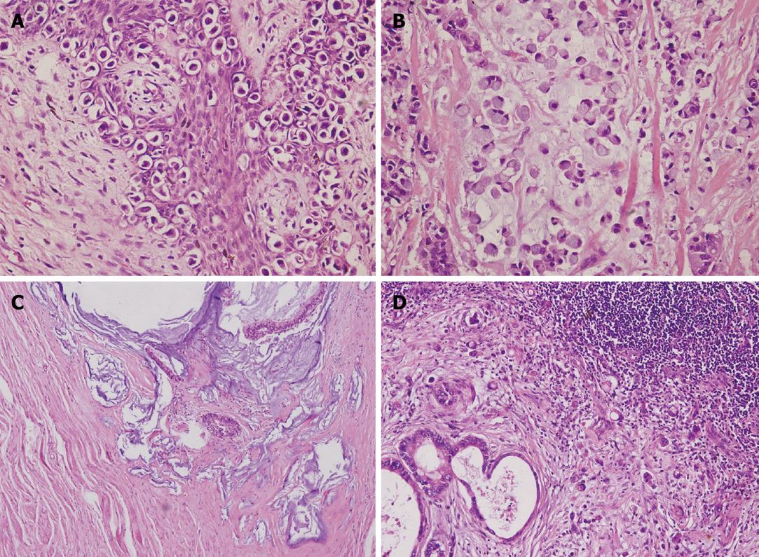
Figure 1 Histopathological images of perianal Paget’s disease, rectal cancer, and metastases in a lymph node from one patient.
A: Paget’s disease under high-power magnification (× 400); B: Signet cell ring cancer component of rectal cancer under high-power magnification (× 400); C: Mucinous adenocarcinoma component of rectal cancer under high-power magnification (× 200); D: Metastases in inguinal lymph node under high-power magnification (× 400).
- Citation: Lian P, Gu WL, Zhang Z, Cai GX, Wang MH, Xu Y, Sheng WQ, Cai SJ. Retrospective analysis of perianal Paget’s disease with underlying anorectal carcinoma. World J Gastroenterol 2010; 16(23): 2943-2948
- URL: https://www.wjgnet.com/1007-9327/full/v16/i23/2943.htm
- DOI: https://dx.doi.org/10.3748/wjg.v16.i23.2943


