Copyright
©2010 Baishideng.
World J Gastroenterol. Jun 14, 2010; 16(22): 2832-2834
Published online Jun 14, 2010. doi: 10.3748/wjg.v16.i22.2832
Published online Jun 14, 2010. doi: 10.3748/wjg.v16.i22.2832
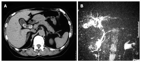
Figure 1 Computed tomography (CT) and magnetic resonance cholangiopancreatography (MRCP).
A: Abdominal CT scan showing 2 stones in the gallbladder and a stone in the common bile duct; B: MRCP revealed a mass (20 mm × 15 mm in diameter) in the dilated common bile duct (20 mm in diameter).
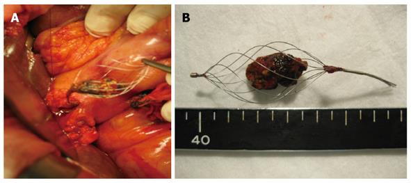
Figure 2 Intraoperative findings.
A: During surgery, the impacted basket with stone were removed through a longitudinal choledochotomy; B: The size of the stone in the basket was 20 mm in diameter.
- Citation: Fukino N, Oida T, Kawasaki A, Mimatsu K, Kuboi Y, Kano H, Amano S. Impaction of a lithotripsy basket during endoscopic lithotomy of a common bile duct stone. World J Gastroenterol 2010; 16(22): 2832-2834
- URL: https://www.wjgnet.com/1007-9327/full/v16/i22/2832.htm
- DOI: https://dx.doi.org/10.3748/wjg.v16.i22.2832


