Copyright
©2010 Baishideng.
World J Gastroenterol. Jan 14, 2010; 16(2): 193-200
Published online Jan 14, 2010. doi: 10.3748/wjg.v16.i2.193
Published online Jan 14, 2010. doi: 10.3748/wjg.v16.i2.193
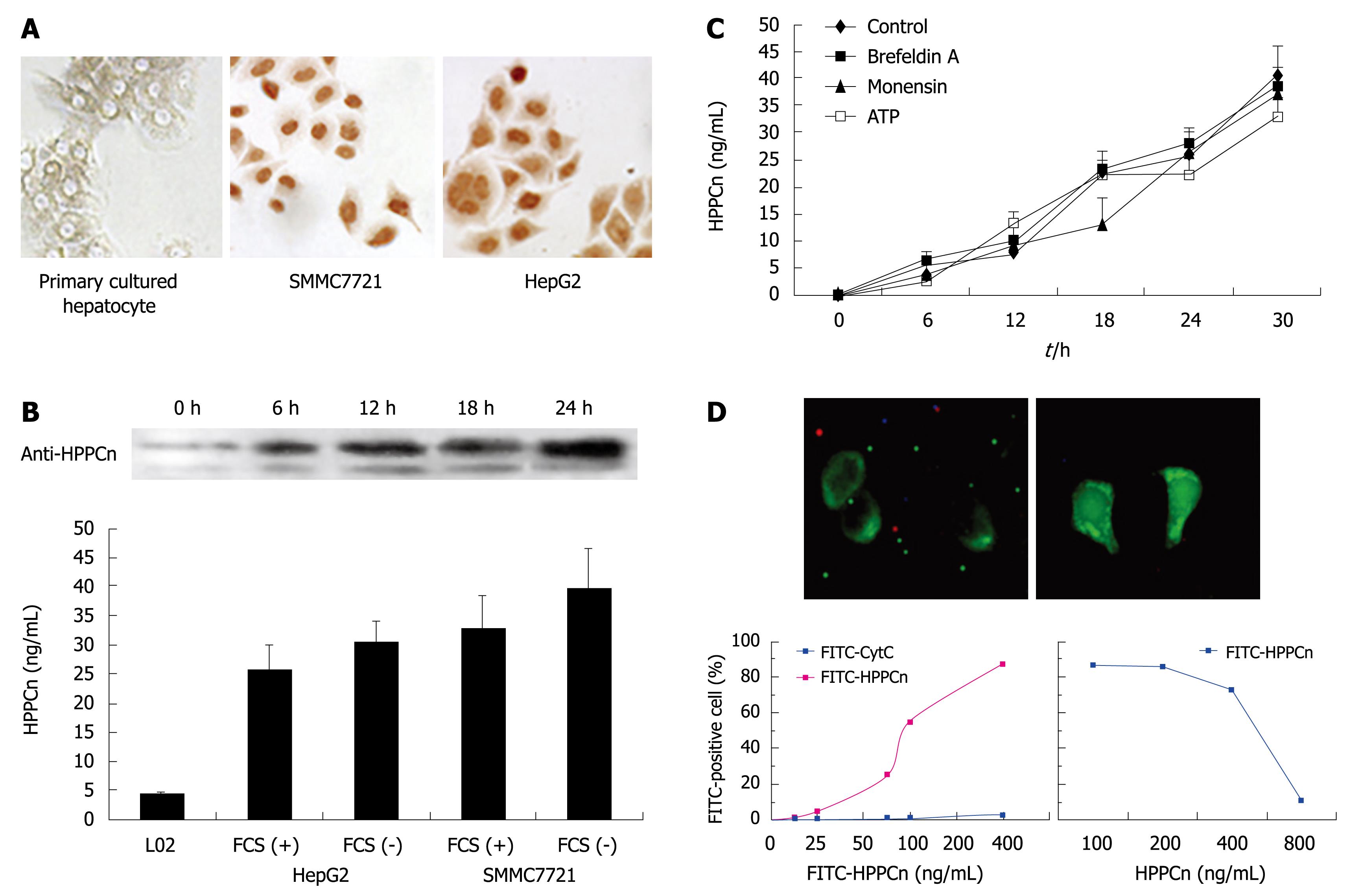
Figure 1 Hepatopoietin Cn (HPPCn) acting as an autocrine factor for hepatocellular carcinoma (HCC) cells in vitro.
A: Immunohistochemistry showing staining with anti-HPPCn antibody in primarily cultured hepatocytes and HCC cells; B: Western blotting and enzyme-linked immunosorbent assay (ELISA) showing the secretion of HPPCn into culture medium (CM) of HCC-derived cells; C: ELISA showing the kinetics of HPPCn secretion from HCC cells; D: Binding of HPPCn to HCC cells.
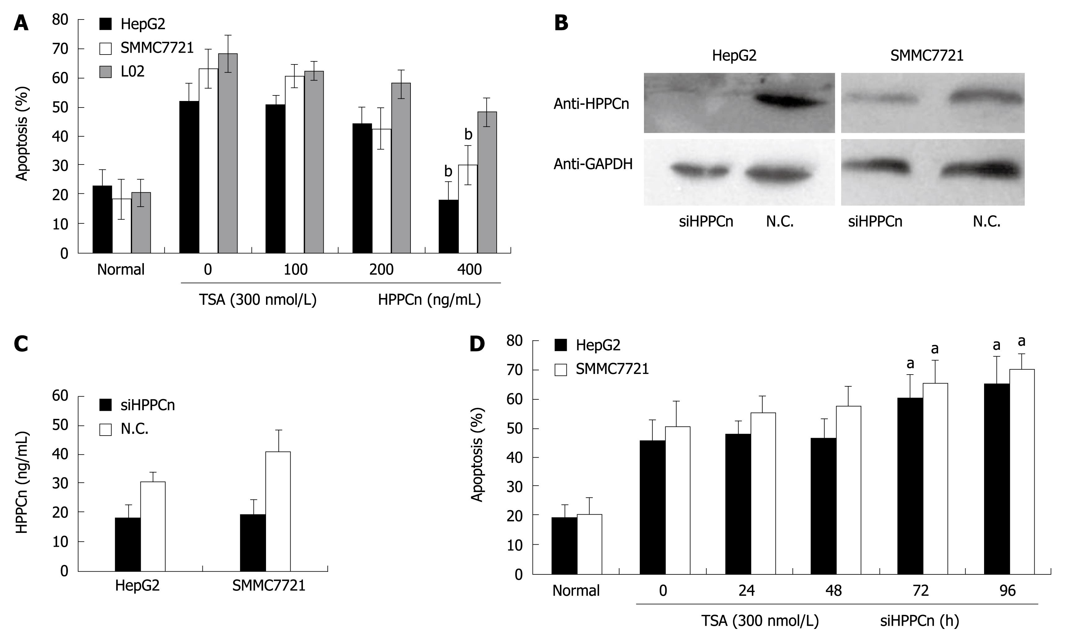
Figure 2 HPPCn suppressing apoptosis of HCC-derived cells.
A: Flow cytometry showing dose-dependent suppressing activity of recombinant human protein (rhHPPCn) on apoptosis of HCC cells incubated with 300 nmol/L trichostatin A (TSA) for 24 h prior to treatment with indicated concentrations of rhHPPCn; B: Western blotting showing hybridization to anti-HPPCn antibody and anti-GAPDH in HepG2 and SMMC7721 transfected with specific siRNA; C: ELISA showing the secretion of HPPCn into CM of HepG2 and SMMC7721; D: Flow cytometry showing the percentages of apoptotic cells incubated with 300 nmol/L TSA for 24 h prior to transfection with specific siRNA for an indicated period of time. The data are expressed as mean ± SD of 3 independent experiments. aP < 0.05, bP < 0.01 vs group without HPPCn/siHPPCn treatment. N.C.: Negative control.
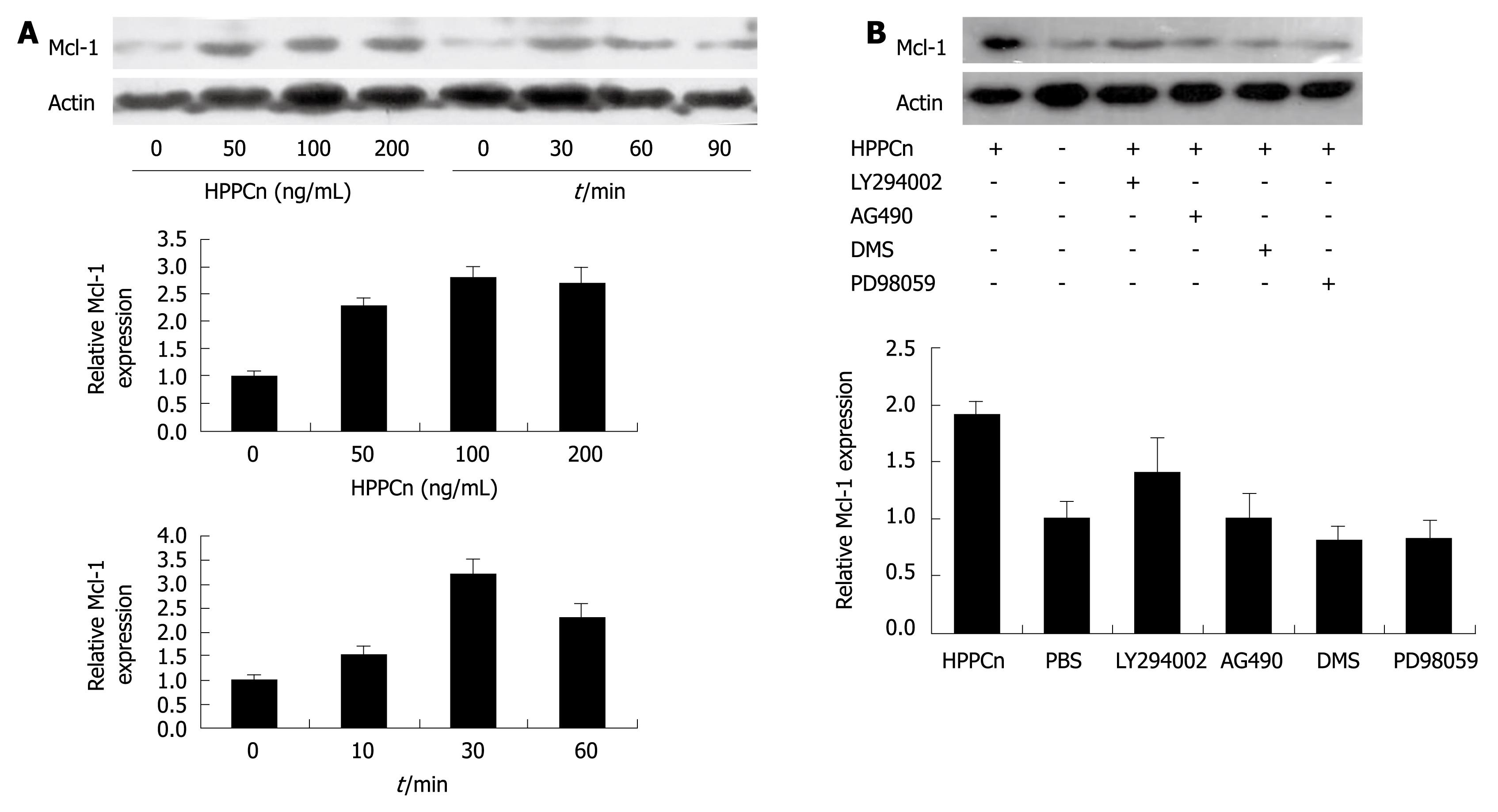
Figure 3 HPPCn inducing Mcl-1 expression via the MEK/ERK and SPK/S1P pathways.
A: HPPCn induces Mcl-1 expression in a dose- and time-dependent manner and the densities of signals were determined by densitometry; B: Western blotting showing hybridization to anti-Mcl-1 antibody and anti-β-actin in HepG2 after the cells were treated with HPPCn for 3 h prior to pre-incubation with DMS, PD98059, LY294002, or AG490 for 1 h.
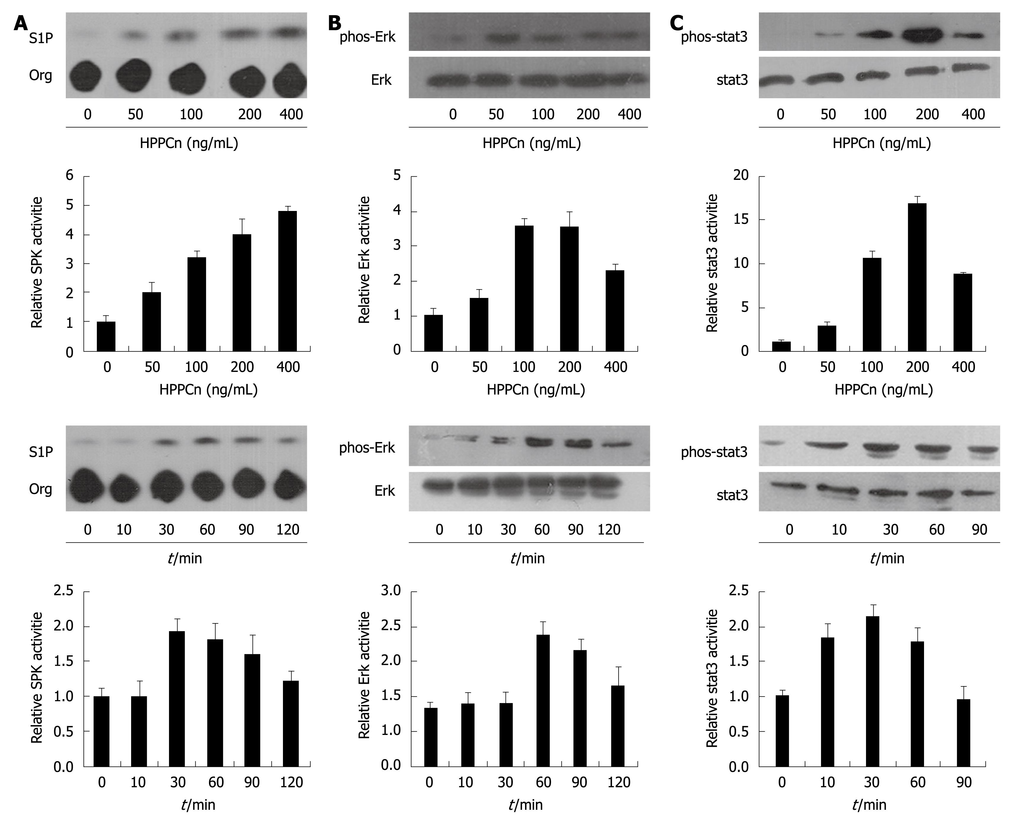
Figure 4 Effects of rhHPPCn on the activities of SPK (A), Jak-Stat3 (B), and Erk1/2 (C).
HepG2 cells were treated for 30 min with the indicated concentration of rhHPPCn and 200 ng/mL rhHPPCn for the indicated periods of time. The phosphorylation of Stat3 and Erk1/2 were determined by Western blotting with antibodies against both species of Stat3 and Erk1/2. The densities of signals were determined by densitometry.
- Citation: Chang J, Liu Y, Zhang DD, Zhang DJ, Wu CT, Wang LS, Cui CP. Hepatopoietin Cn suppresses apoptosis of human hepatocellular carcinoma cells by up-regulating myeloid cell leukemia-1. World J Gastroenterol 2010; 16(2): 193-200
- URL: https://www.wjgnet.com/1007-9327/full/v16/i2/193.htm
- DOI: https://dx.doi.org/10.3748/wjg.v16.i2.193


