Copyright
©2010 Baishideng.
World J Gastroenterol. May 7, 2010; 16(17): 2190-2192
Published online May 7, 2010. doi: 10.3748/wjg.v16.i17.2190
Published online May 7, 2010. doi: 10.3748/wjg.v16.i17.2190
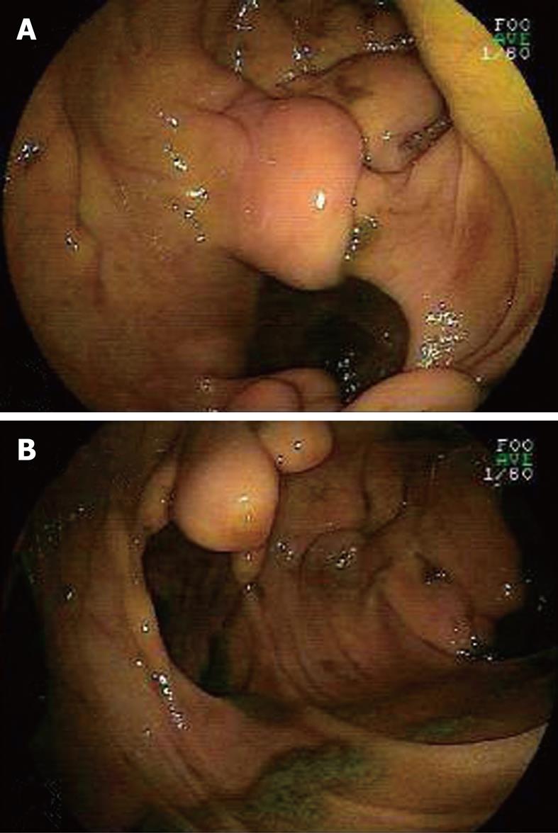
Figure 1 Double-balloon endoscopy (DBE) by the oral approach revealing several rounded protuberances in the jejunum (B) and a diverticulum-like hole (A).
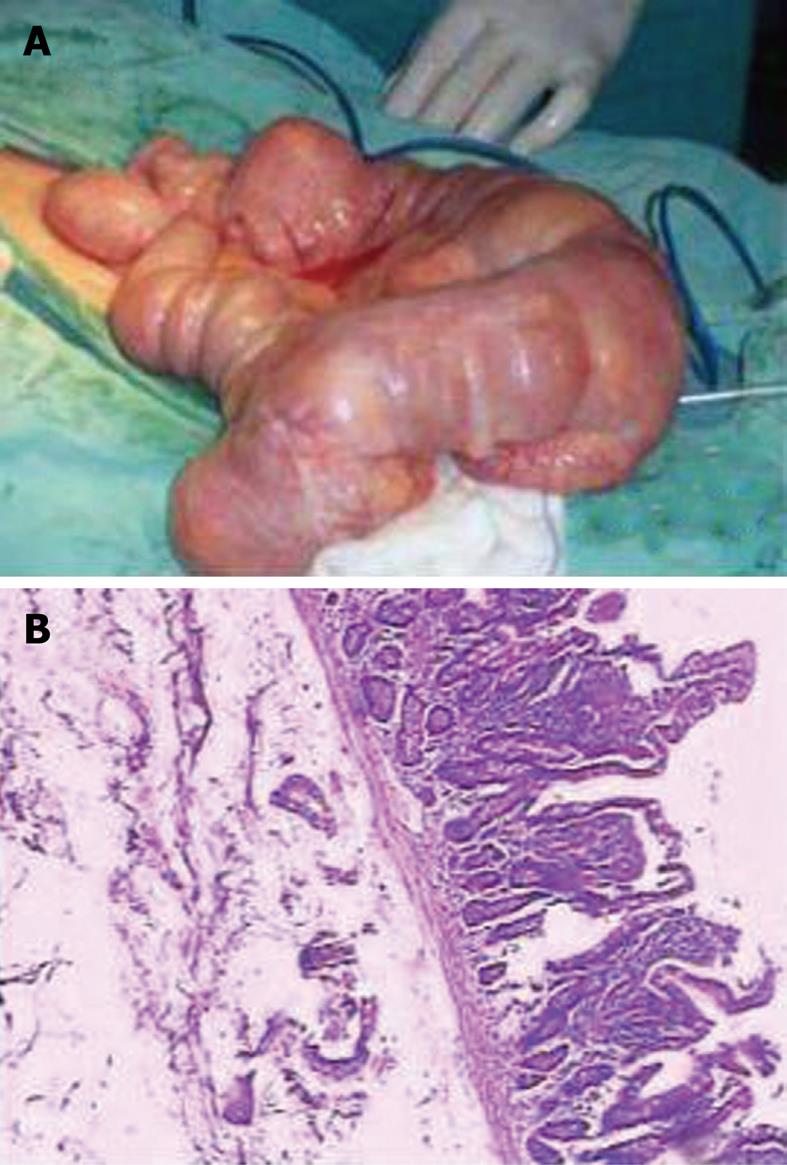
Figure 2 Resected specimen of jejunal tissue.
A: Macroscopic appearance of the resected specimen with seven remarkably dilated cysts; B: Microscopic appearance of resected tissue showing multiple lipomas lining within intestinal wall (HE, × 20).
- Citation: Wan XY, Deng T, Luo HS. Partial intestinal obstruction secondary to multiple lipomas within jejunal duplication cyst: A case report. World J Gastroenterol 2010; 16(17): 2190-2192
- URL: https://www.wjgnet.com/1007-9327/full/v16/i17/2190.htm
- DOI: https://dx.doi.org/10.3748/wjg.v16.i17.2190


