Copyright
©2010 Baishideng.
World J Gastroenterol. May 7, 2010; 16(17): 2187-2189
Published online May 7, 2010. doi: 10.3748/wjg.v16.i17.2187
Published online May 7, 2010. doi: 10.3748/wjg.v16.i17.2187
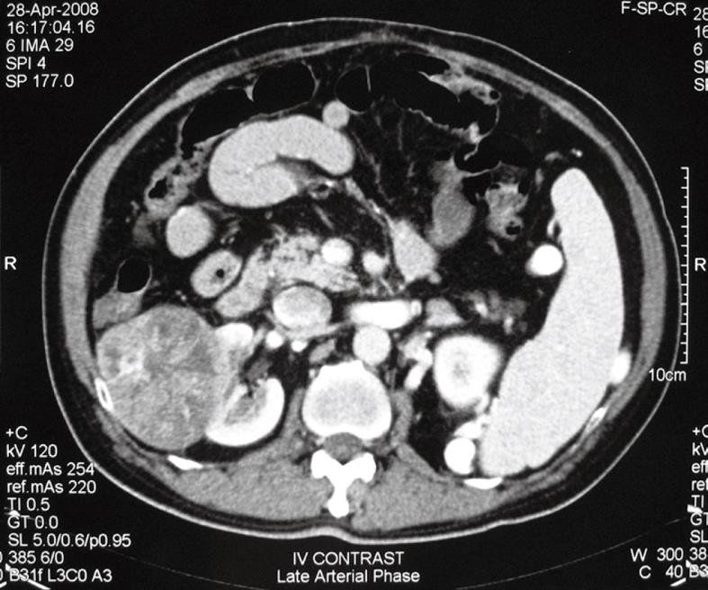
Figure 1 Computed tomography showing the contrast-enhanced mass in the right kidney.
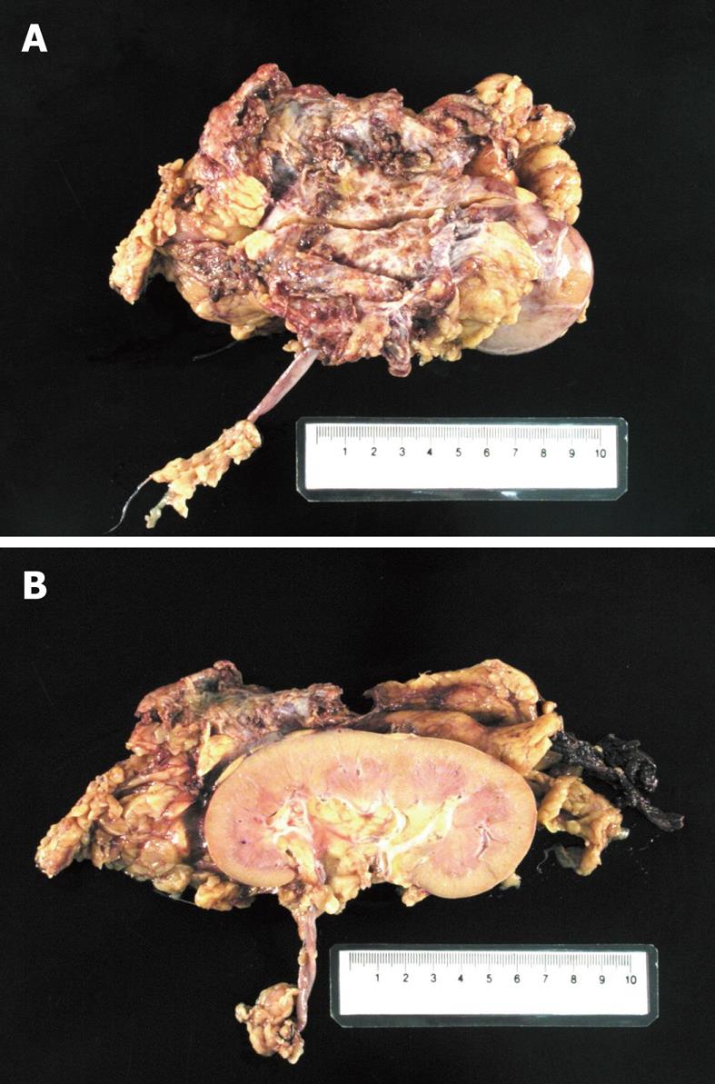
Figure 2 Gross specimen of tumor showing its relationship with the right kidney.
A: External appearance; B: From cut section of kidney.
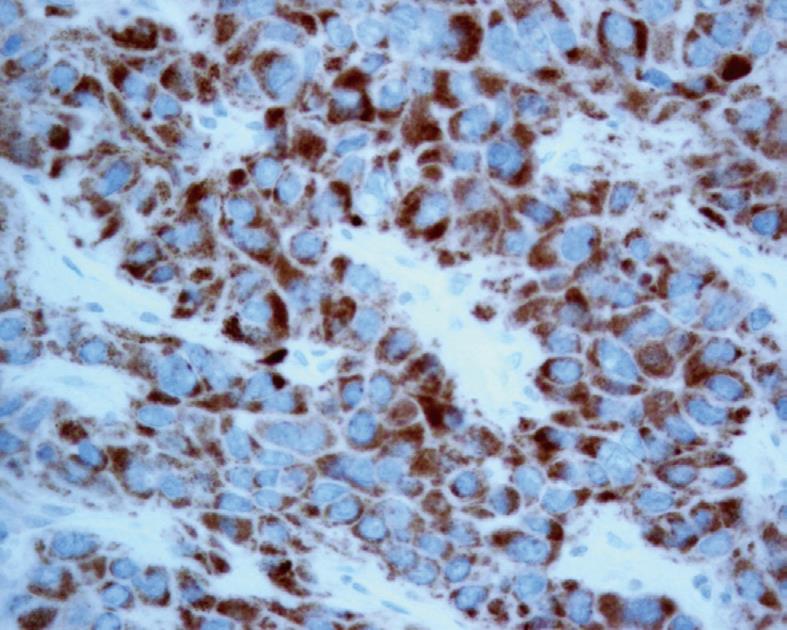
Figure 3 Immunological stain of the tumor with HepPar, confirmed the tumor as hepatocellular carcinoma.
- Citation: Wong TCL, To KF, Hou SSM, Yip SKH, Ng CF. Late retroperitoneal recurrence of hepatocellular carcinoma 12 years after initial diagnosis. World J Gastroenterol 2010; 16(17): 2187-2189
- URL: https://www.wjgnet.com/1007-9327/full/v16/i17/2187.htm
- DOI: https://dx.doi.org/10.3748/wjg.v16.i17.2187


