Copyright
©2009 The WJG Press and Baishideng.
World J Gastroenterol. Mar 7, 2009; 15(9): 1144-1146
Published online Mar 7, 2009. doi: 10.3748/wjg.15.1144
Published online Mar 7, 2009. doi: 10.3748/wjg.15.1144
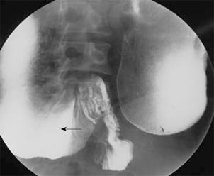
Figure 1 Barium image from upper gastrointestinal series reveals duodenal obstruction, demonstrating high-grade stenosis of the fourth portion of the duodenum and extreme dilation of proximal duodenum (arrow).
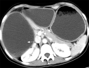
Figure 2 CT scan shows duodenum does not cross behind the superior mesenteric artery and the superior mesenteric vein, and extreme dilation of proximal duodenum (arrow).
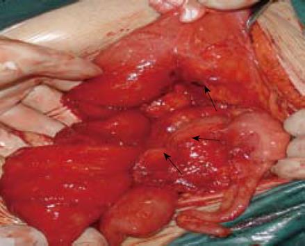
Figure 3 One band running from the anti-mesenteric wall of proximal jejunum to cecum has been lysed (arrows) and no Treitz’s ligament is found.
- Citation: Gong J, Zheng ZJ, Mai G, Liu XB. Malrotation causing duodenal chronic obstruction in an adult. World J Gastroenterol 2009; 15(9): 1144-1146
- URL: https://www.wjgnet.com/1007-9327/full/v15/i9/1144.htm
- DOI: https://dx.doi.org/10.3748/wjg.15.1144


