Copyright
©2009 The WJG Press and Baishideng.
World J Gastroenterol. Mar 7, 2009; 15(9): 1105-1112
Published online Mar 7, 2009. doi: 10.3748/wjg.15.1105
Published online Mar 7, 2009. doi: 10.3748/wjg.15.1105
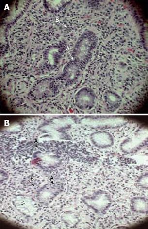
Figure 1 The histopathological features of active gastritis in DU and TR.
A: Polymorphonuclear cells infiltration inside glands (HE, × 400) (A) and in lamina propria (B)-DU (HE, × 400); B: Neutrophils in glands (A) (HE, × 400) and in lamina propria (B)-TR (HE, × 400).
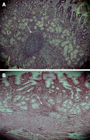
Figure 2 Mononuclear cells, lymphoid follicles and aggregates in DU and TR.
A: Arrow indicates mononuclear cells in lamina propria with a lymphoid follicle in DU (HE, × 200); B: Arrow indicates mononuclear cells in lamina propria with lymphoid aggregate in TR (HE, × 200).
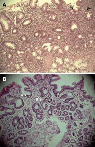
Figure 3 Intestinal metaplastic changes in DU and TR.
A: Metaplastic changes with goblet cells in glands (B) and surface epithelium of gastric mucosa (A)-DU (HE, × 200); B: Metaplastic cells replacing the normal gastric epithelium in glands (B) and surface mucosal epithelium (A)-TR (HE, × 200).
-
Citation: Saha DR, Datta S, Chattopadhyay S, Patra R, De R, Rajendran K, Chowdhury A, Ramamurthy T, Mukhopadhyay AK. Indistinguishable cellular changes in gastric mucosa between
Helicobacter pylori infected asymptomatic tribal and duodenal ulcer patients. World J Gastroenterol 2009; 15(9): 1105-1112 - URL: https://www.wjgnet.com/1007-9327/full/v15/i9/1105.htm
- DOI: https://dx.doi.org/10.3748/wjg.15.1105


