Copyright
©2009 The WJG Press and Baishideng.
World J Gastroenterol. Feb 28, 2009; 15(8): 912-918
Published online Feb 28, 2009. doi: 10.3748/wjg.15.912
Published online Feb 28, 2009. doi: 10.3748/wjg.15.912
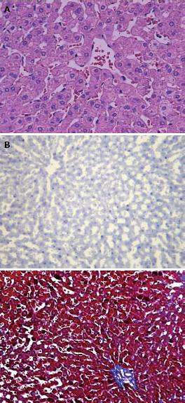
Figure 1 The rabbits fed with the standard diet.
A: Normal liver cells without vacuolar degeneration and no distinct inflammatory cell infiltration by HE staining (× 400); B: No positive fat deposition by red-oil staining (× 100); C: Normal liver cells without apparent collagen fibrosis by Masson trichrome staining (× 100).
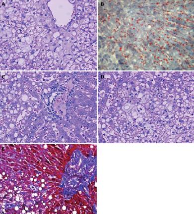
Figure 2 The rabbits fed with the high-fat diet.
A: Pronounced hepatic steatosis. Hepatocellular ballooning with clear vacuolar degeneration was apparent by HE staining (× 400); B: Pronounced hepatic steatosis with positive fat infiltration by red-oil staining (× 200); C: Abundant mononuclear and polymorphonuclear inflammatory cells infiltrates by HE staining (× 400); D: Pieces necrosis by HE staining (× 400); E: Significant collagen and reticulin fibrosis among hepatic cells in blue color by Masson trichrome staining (× 200).
- Citation: Fu JF, Fang YL, Liang L, Wang CL, Hong F, Dong GP. A rabbit model of pediatric nonalcoholic steatohepatitis: The role of adiponectin. World J Gastroenterol 2009; 15(8): 912-918
- URL: https://www.wjgnet.com/1007-9327/full/v15/i8/912.htm
- DOI: https://dx.doi.org/10.3748/wjg.15.912


