Copyright
©2009 The WJG Press and Baishideng.
World J Gastroenterol. Feb 21, 2009; 15(7): 882-884
Published online Feb 21, 2009. doi: 10.3748/wjg.15.882
Published online Feb 21, 2009. doi: 10.3748/wjg.15.882
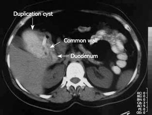
Figure 1 Oral and intravenous contrast-enhanced abdominal CT imaging.
The nasogastric tube was extended into the cyst, which was filled with oral contrast medium, through the defect between the duodenum and the cyst.
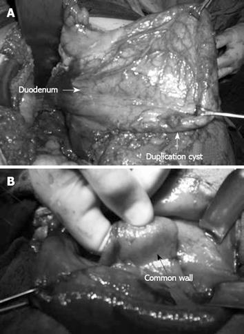
Figure 2 Operative view of cyst.
A: Duodenal duplication cyst; B: Inner surface of the cyst.
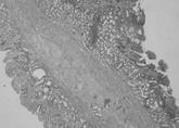
Figure 3 Common wall containing double mucosa with muscularis mucosa on each side and intervening connective tissue fibers (Hematoxylin and eosin, × 10).
- Citation: Uzun MA, Koksal N, Kayahan M, Celik A, Kılıcoglu G, Ozkara S. A rare case of duodenal duplication treated surgically. World J Gastroenterol 2009; 15(7): 882-884
- URL: https://www.wjgnet.com/1007-9327/full/v15/i7/882.htm
- DOI: https://dx.doi.org/10.3748/wjg.15.882


