Copyright
©2009 The WJG Press and Baishideng.
World J Gastroenterol. Feb 21, 2009; 15(7): 879-881
Published online Feb 21, 2009. doi: 10.3748/wjg.15.879
Published online Feb 21, 2009. doi: 10.3748/wjg.15.879
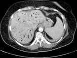
Figure 1 Extensive gas accumulation in the hepatic-portal vein branches.
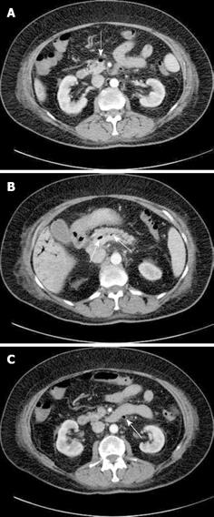
Figure 2 Gas observed.
A: Superior mesenteric vein (arrow); B: Splenic vein (arrows); C: Inferior mesenteric vein (arrow).
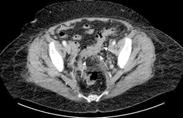
Figure 3 Diverticulosis in the sigmoid colon and mild inflammatory changes are present in the sigmoid mesocolon (arrows).
- Citation: Şen M, Akpınar A, İnan A, Şişman M, Dener C, Akın K. Extensive hepatic-portal and mesenteric venous gas due to sigmoid diverticulitis. World J Gastroenterol 2009; 15(7): 879-881
- URL: https://www.wjgnet.com/1007-9327/full/v15/i7/879.htm
- DOI: https://dx.doi.org/10.3748/wjg.15.879


