Copyright
©2009 The WJG Press and Baishideng.
World J Gastroenterol. Feb 7, 2009; 15(5): 599-606
Published online Feb 7, 2009. doi: 10.3748/wjg.15.599
Published online Feb 7, 2009. doi: 10.3748/wjg.15.599
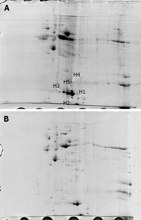
Figure 1 Differential two-dimensional maps of wild-type strain Hp27 (A) and mutant strain Hp27_ΔcagA (B) stained with Commassie blue.
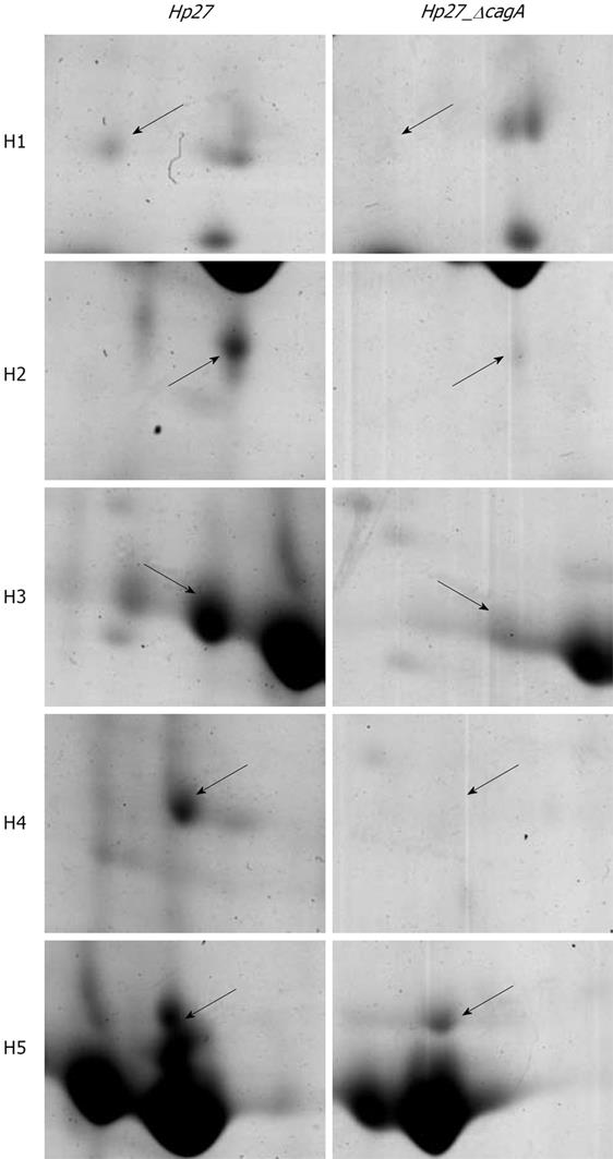
Figure 2 Magnified segments of 2-DE gel map of protein spots including H1~H5.
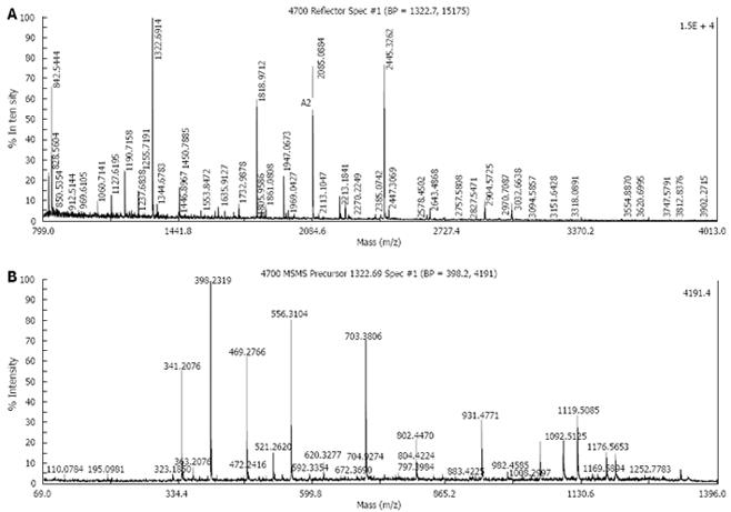
Figure 3 Mass spectrometry (MS) maps of one protein spots.
A: MS of H1; B: MS/MS of H1.
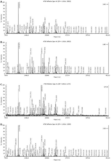
Figure 4 Mass spectrometry (MS) maps of 4 protein spots.
A: MS of H2; B: MS of H3; C: MS of H4;
-
Citation: Huang ZG, Duan GC, Fan QT, Zhang WD, Song CH, Huang XY, Zhang RG. Mutation of cytotoxin-associated gene A affects expressions of antioxidant proteins of
Helicobacter pylori . World J Gastroenterol 2009; 15(5): 599-606 - URL: https://www.wjgnet.com/1007-9327/full/v15/i5/599.htm
- DOI: https://dx.doi.org/10.3748/wjg.15.599


