Copyright
©2009 The WJG Press and Baishideng.
World J Gastroenterol. Dec 14, 2009; 15(46): 5799-5804
Published online Dec 14, 2009. doi: 10.3748/wjg.15.5799
Published online Dec 14, 2009. doi: 10.3748/wjg.15.5799
Figure 1 Spontaneous electrical activity of smooth muscle cells in human colon and small intestine.
A: Colonic smooth muscle cells produced slow waves with a frequency of 18.1 ± 2.1/min; B: Small intestinal smooth muscle cells also produced slow waves with a frequency of 3.1 ± 0.5/min.
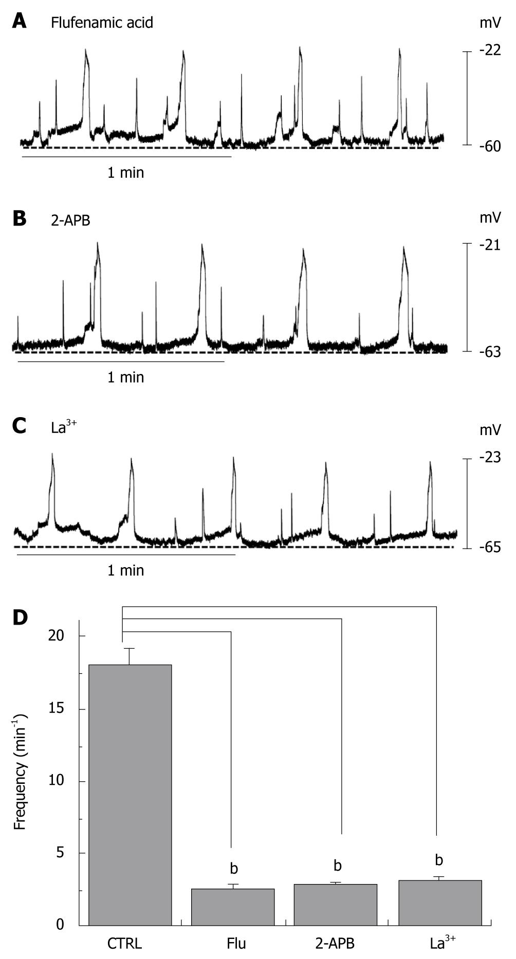
Figure 2 Effects of flufenamic acid, 2-aminoethoxydiphenyl borate (2-APB) and La3+ on electrical responses.
Flufenamic acid (50 μmo/L, A), 2-APB (50 μmo/L, B) or La3+ (50 μmo/L, C) were applied while recording electrical activity of isolated smooth muscles of the human colon. All drugs inhibited the spontaneous electrical activity; D: The histograms summarize the frequency of spontaneous electrical activities in human colon with flufenamic acid, 2-APB, and La3+. bP < 0.01.
Figure 3 Expression of transient receptor potential melastatin-type 7 (TRPM7) protein in human colon.
Double labeling of TRPM7-like immunoreactivity (red) and c-kit–like immunoreactivity (green) within smooth muscle layers of the human colon. The mixed color yellow indicates the colocalization of both TRPM7-like and c-kit–like immunoreactivity (bar = 50 μm). DIC: differential interference contrast.
Figure 4 Expression of TRPM7 protein in human small intestine.
Double labeling of TRPM7-like immunoreactivity (red) and c-kit–like immunoreactivity (green) within smooth muscle layers of human small intestine. The mixed color yellow indicates the colocalization of both TRPM7-like and c-kit–like immunoreactivity (bar = 50 μm).
- Citation: Kim BJ, Park KJ, Kim HW, Choi S, Jun JY, Chang IY, Jeon JH, So I, Kim SJ. Identification of TRPM7 channels in human intestinal interstitial cells of Cajal. World J Gastroenterol 2009; 15(46): 5799-5804
- URL: https://www.wjgnet.com/1007-9327/full/v15/i46/5799.htm
- DOI: https://dx.doi.org/10.3748/wjg.15.5799


