Copyright
©2009 The WJG Press and Baishideng.
World J Gastroenterol. Dec 14, 2009; 15(46): 5770-5775
Published online Dec 14, 2009. doi: 10.3748/wjg.15.5770
Published online Dec 14, 2009. doi: 10.3748/wjg.15.5770
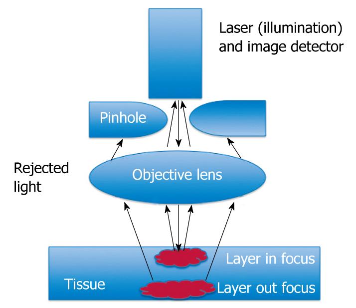
Figure 1 Schematic of confocal endomicroscopy principles.
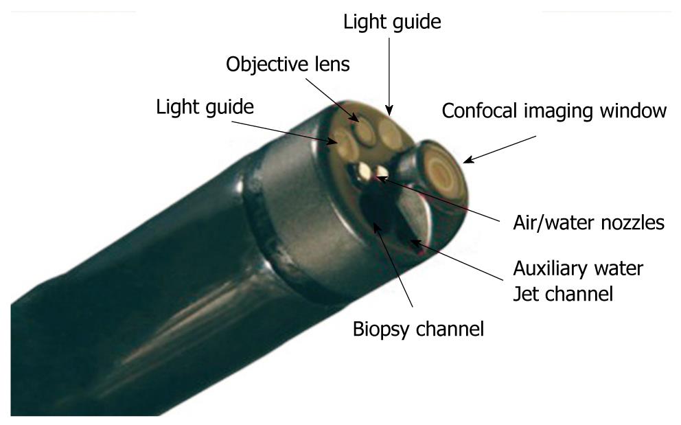
Figure 2 The endoscope-based confocal laser endomicroscopy (eCLE) imaging system: the distal tip.
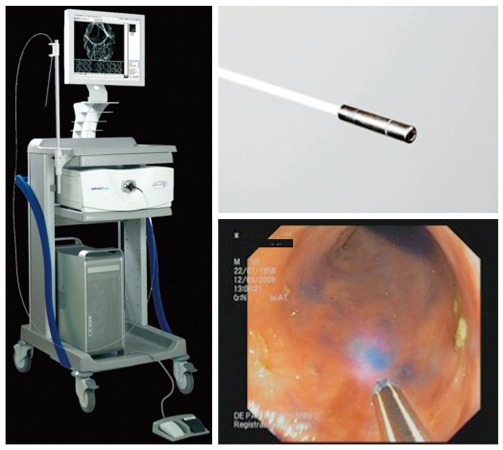
Figure 3 The probe-based CLE (pCLE) imaging system.
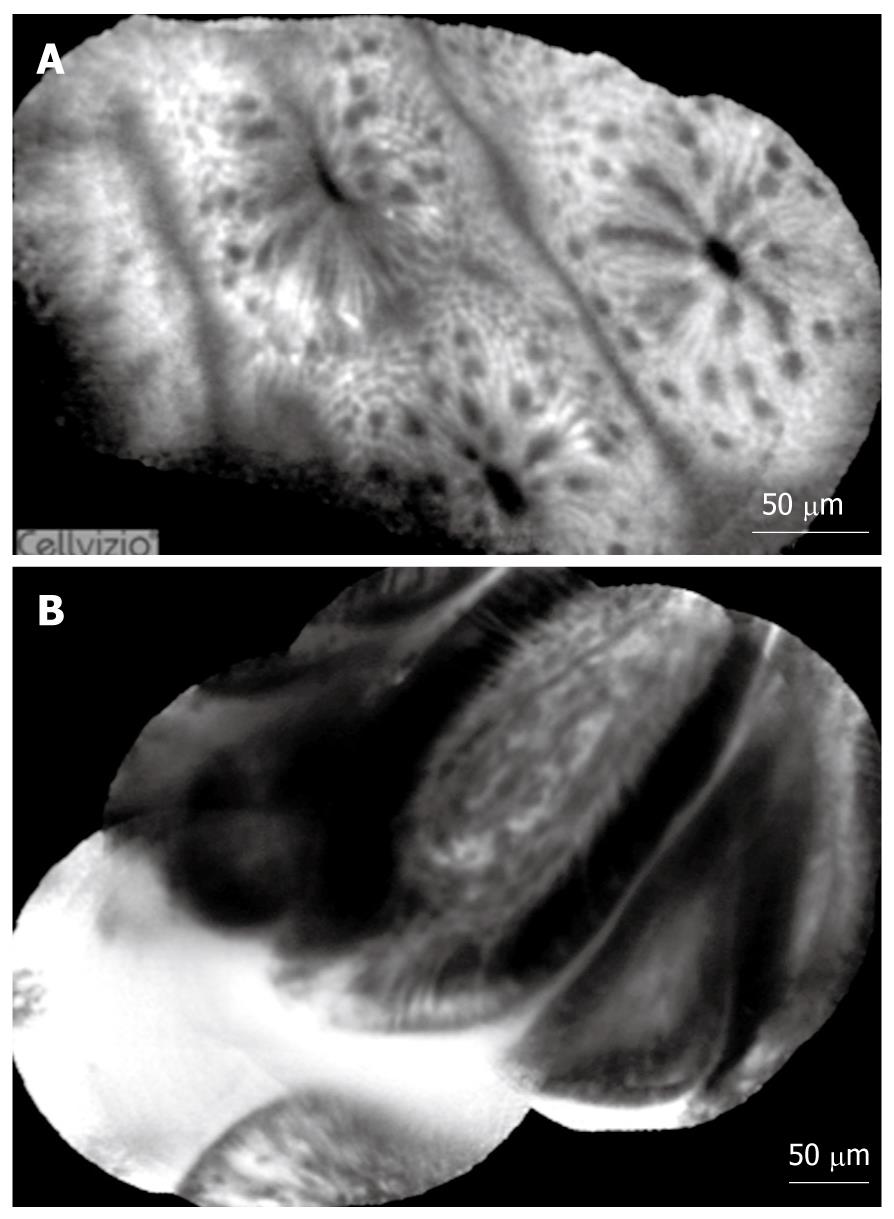
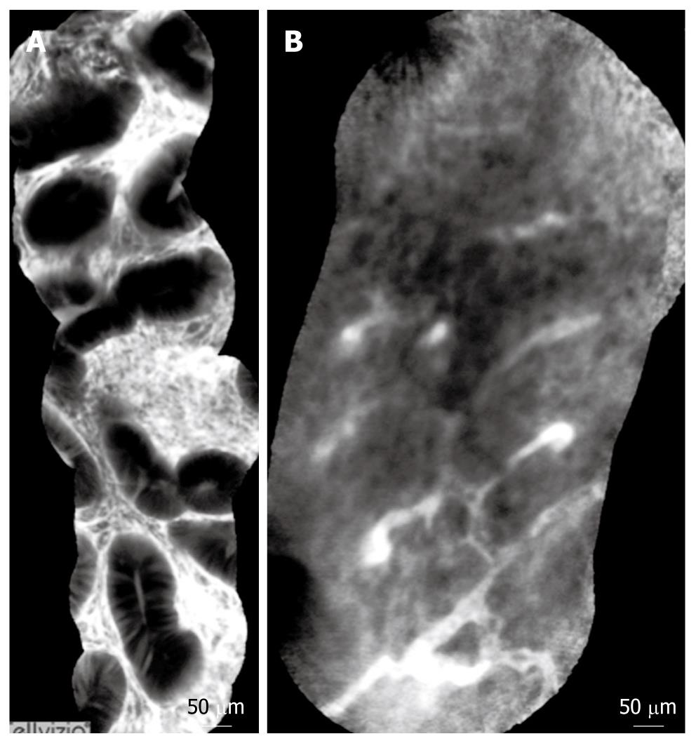
Figure 4 pCLE fluorescein sodium 10% imaging of the normal colon (A) and normal duodenum (B).
Figure 5 pCLE fluorescein sodium 10% imaging of an adenomatous colonic polyp (A) and colonic adenocarcinoma (B).
-
Citation: Palma GDD. Confocal laser endomicroscopy in the “
in vivo ” histological diagnosis of the gastrointestinal tract. World J Gastroenterol 2009; 15(46): 5770-5775 - URL: https://www.wjgnet.com/1007-9327/full/v15/i46/5770.htm
- DOI: https://dx.doi.org/10.3748/wjg.15.5770


